Sectra Implant Movement Analysis
Product | Orthopaedics
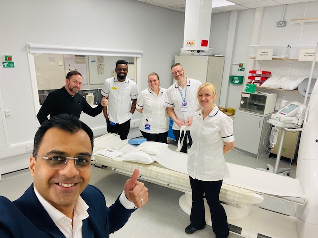
 Wrightington, Wigan & Leigh Teaching Hospitals NHS Foundation
Radiologists, radiographers and surgeons at Wrightington, Wigan & Leigh Teaching Hospitals have been the first in the UK to assess Sectra IMA – a dynamic CT technique giving clearer insight into implant loosening, reducing unnecessary revision surgery, and enhancing outcomes. Dr Subhasis Basu and Karen Haworth explain the potential of an initiative that could save significant costs for the NHS and improve patients’ lives.
Wrightington, Wigan & Leigh Teaching Hospitals NHS Foundation Trust is no stranger to surgical innovation.
Sir John Charnley famously performed the world’s first total hip replacement at Wrightington Hospital in 1962: one of many significant contributions to orthopaedic practice.
Still one of the highest volume centres for primary and revision arthroplasty anywhere in the UK, WWL, as the trust is widely known, continues to be recognised for its commitment to modernising surgical approaches.
With that, pioneering healthcare professionals at the trust have become the first in the UK to evaluate a diagnostic technique that has the potential to redefine how a set of costly and potentially harmful post-operative complications are assessed, identified, and acted on.
Particularly when patients present with pain following hip and knee replacements, radiologists, radiographers and surgeons have been exploring a new way to remove uncertainty and to accurately determine whether implants have loosened, and if revision surgery is appropriate.
For approximately 70% of cases, x-rays can be used to indicate implant loosening and to determine the correct clinical response. But for a remaining 30% of patients, the question of implant loosening can remain inconclusive, with little insight to drive clinical decisions on the best course of action.
“Delayed diagnosis of implant loosening can lead to delayed surgery which in turn can lead to patient deterioration and more complex surgery, whilst unnecessary surgery can consume significant resource and risk avoidable harm,” says Dr Subhasis Basu, a consultant musculoskeletal radiologist, who is leading the initiative.
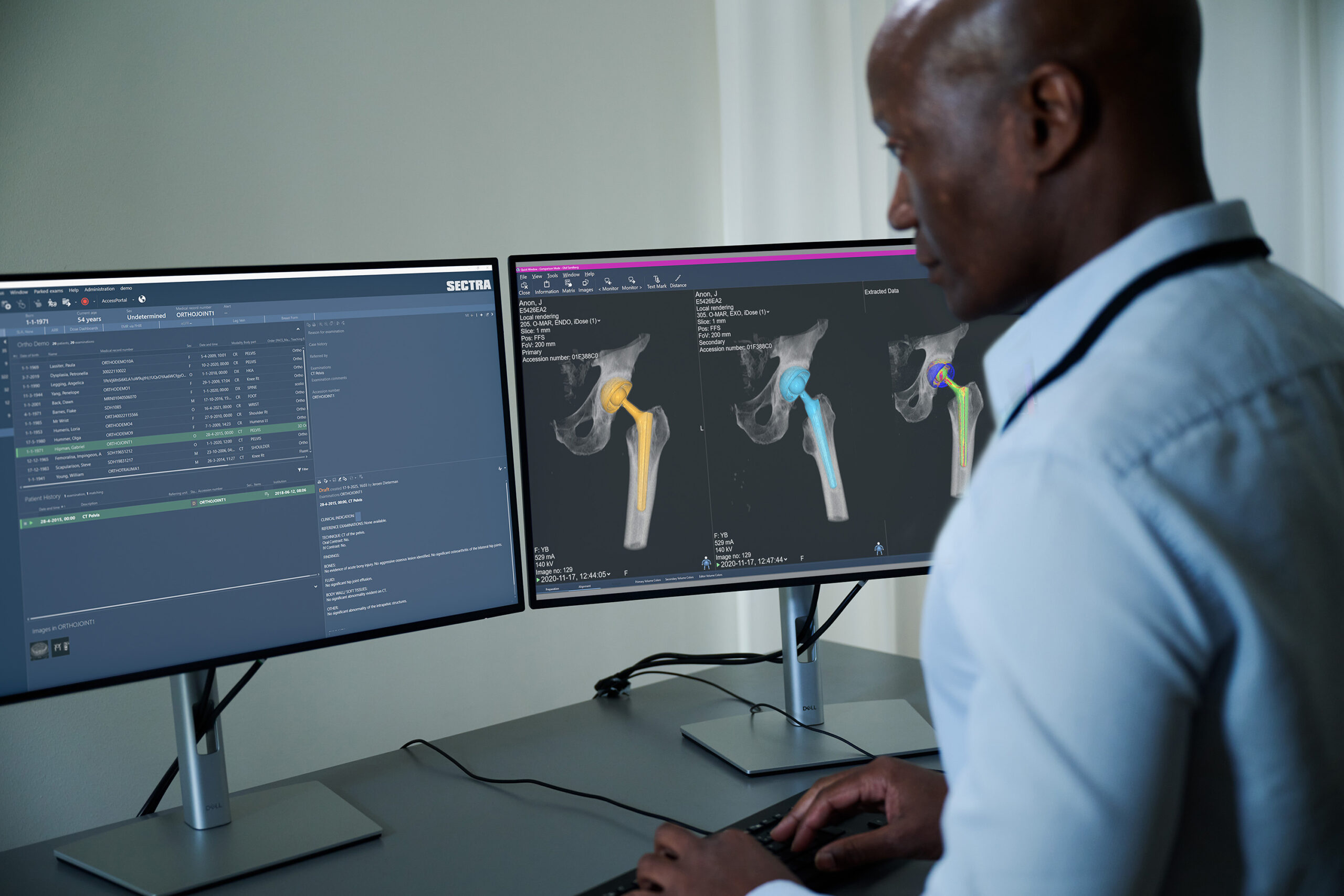
WWL is the first NHS organisation to assess a novel use of dynamic CT to more accurately identify if loosening is happening or not. Its name: Sectra Implant Movement Analysis (IMA).
“We are examining if this is a means to explore implant loosening in a way we have never been able to do before,” says Dr Basu.
“If we can detect loosening earlier and more dynamically than serial static imaging allows – using a single study performed at a single point in time – that gives us the ability to assess loosening more effectively. That is very helpful diagnostically and helps the patient in their pathway to potentially reach an earlier diagnosis.”
Dr Basu notes that demand for primary hip and knee surgery is increasing both across the UK and worldwide. With individual revision procedures costing as much as £100,000, particularly if they are complex, also carrying risks of additional complications, IMA offers the potential to identify the right patients for the right pathways.
“The key point is trying to identify quite early in that pathway when somebody is more suitable for a revision and when somebody isn’t,” he says.
“We often see patients coming back and forth into clinics, having repeat follow up x-rays to compare studies and the appearances of implant positions, often to find no significant change. It’s an ever-repetitive cycle.”
“There is the potential that IMA can really help break that cycle,” he says. “To have that bit of information that could really identify and provide further diagnostic information beyond the conventional static imaging, could then help make that key decision at a key moment in time, that doesn’t otherwise transpire.”
IMA isn’t suitable for every patient, he adds, explaining its key value is when other imaging remains inconclusive and the patient is still symptomatic. “It’s about choosing the right patients where the results can genuinely influence clinical decision-making. With rising imaging volumes and stretched radiology capacity, we have to be smarter about who we image, how and when. For a select subset of patients, IMA offers a level of diagnostic insight we simply didn’t have before.”
He believes IMA could provide an “important number of patients” with a “decision that works in their favour.” NHS teams could identify if an implant is loose (or not), as well as having deeper insights into the extent of loosening, informing both the need for surgery, and more detailed insights for surgeons when it comes to pre-operative planning.
“All of this could have a significant impact on waiting lists up and down the country given the high numbers of primary and revision procedures that are being performed right now,” says Dr Basu.
It has had an impact in a short time. Surgeons have been able to better explain to patients when and why revision surgery may not necessarily be imminent, because the studies can be more reassuring whilst patients are performing well through their activities of daily living.
It is always an important saying in our clinical practice to ‘treat the man, not the scan’ i.e. treat the patients clinically rather than based on imaging alone. IMA is that added piece in the diagnostic jigsaw puzzle that contributes to better delivery of care.
“Everything boils down to the individual patients and their needs,” says Dr Basu, explaining IMA has the potential to provide further information before the informed consent process begins.
“Not everything is black and white in radiology,” he says. “In fact, it’s different shades of grey.”
“If imaging demonstrates an abnormality, but then the patient is functioning at a reasonably good level – e.g. going to the gym, gardening, walking many miles and able to enjoy daily activities of living in a relatively comfortable way, you don’t create more harm than is necessary by carrying out additional and/or un-necessary surgery. If the IMA study demonstrates no abnormal implant movement, then surgery may not be imminently indicated, or at the very least could be delayed for the patient.
“The flip side is you don’t want what could be a very simple revision surgery to convert into a potentially more complex one because we’ve sat on something for a while where greater loosening and bone loss has occurred.”
IMA informs that decision. “It’s a combination of information gathering, detective work, and then putting it all together for that individual patient,” he says. “Those patients might be treated very differently based on their individual circumstances. But our surgeons will be able to provide more clarity with added insight, working collaboratively through our multidisciplinary practice which is also crucial to highlight when using IMA.”
Radiographers have played a key role in the trust being able to assess IMA, to introduce it to patients, and acquire the quality of images required. “The global pioneer of this actually stated that these are some of the best IMA images he’s ever seen, and kudos to the skill and talent of our WWL radiographers who were quick to grasp the concepts around IMA image acquisition and implement it so well practically,” says Dr Basu.
Radiographers underwent specialist training from Sectra to understand how to position patients and capture required images.
Karen Haworth, deputy superintendent radiographer for the trust, said explanation to patients has been key. “It’s not every day you get to be the first to do something in the UK. The patients were quite excited when they were told that they were the first in the country to undergo this scan. It has all gone really well.”
“Patients are usually in quite a bit of pain as a result of their condition,” she says. “So then explaining to them they may potentially have slightly more discomfort with the position, can make them a little unsure.
“But once we actually do it, many remark it’s not as bad as they thought it was going to be.”
Scans take around 10 minutes to complete. Radiographers use positioning aids to help patients remain still. Hips are turned inwards and outwards by radiographers to show the implant in different positions.
“The patients don’t seem to struggle with the positions. A few said that it was a little bit uncomfortable, but it was bearable. The more they can tolerate it, the better the outcome is going to be. If patients know they might not need a revision as a result, they are all for it.”
Surgeons have already found insights from IMA valuable in their decisions and conversations with patients, says Dr Basu. “It has had an impact in a short time. Surgeons have been able to better explain to patients when and why revision surgery may not necessarily be imminent, because the studies can be more reassuring whilst patients are performing well through their activities of daily living.
“It is always an important saying in our clinical practice to ‘treat the man, not the scan’ i.e. treat the patients clinically rather than based on imaging alone. IMA is that added piece in the diagnostic jigsaw puzzle that contributes to better delivery of care.”
His aim from evidence being gathered: “To see early, positive outcomes in terms of real-life changes for a set of patients with total hip replacements and total knee replacements that have unexplained symptoms. And being able to make diagnoses earlier to help institute appropriate treatment sooner.”