

Standardization in ophthalmic imaging
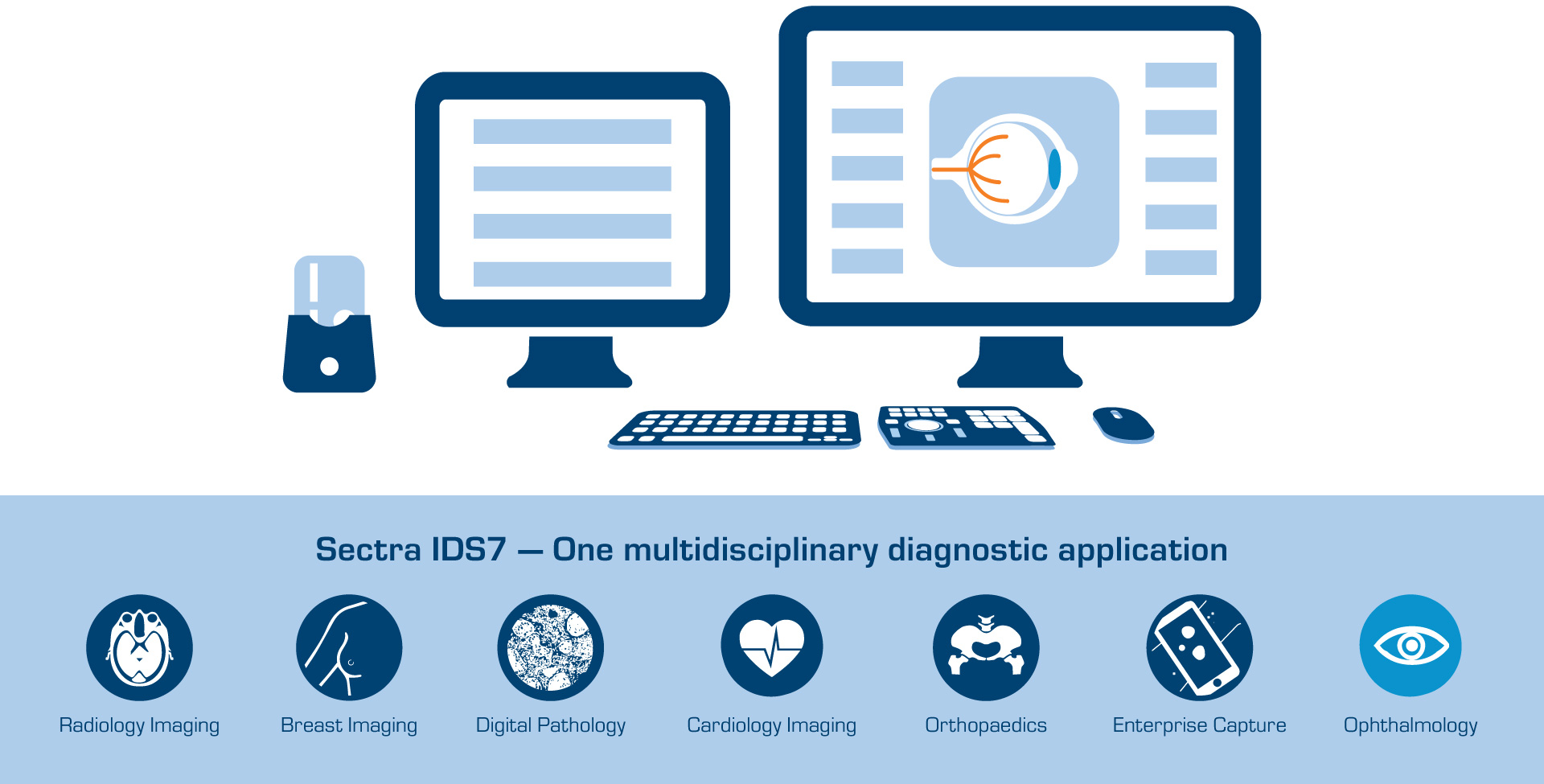
Standardization is key to interoperability across systems and devices. This allows for brilliant workflows, more efficient patient care, easier image sharing, as well as enabling the creation of comprehensive datasets for research and AI analyses.
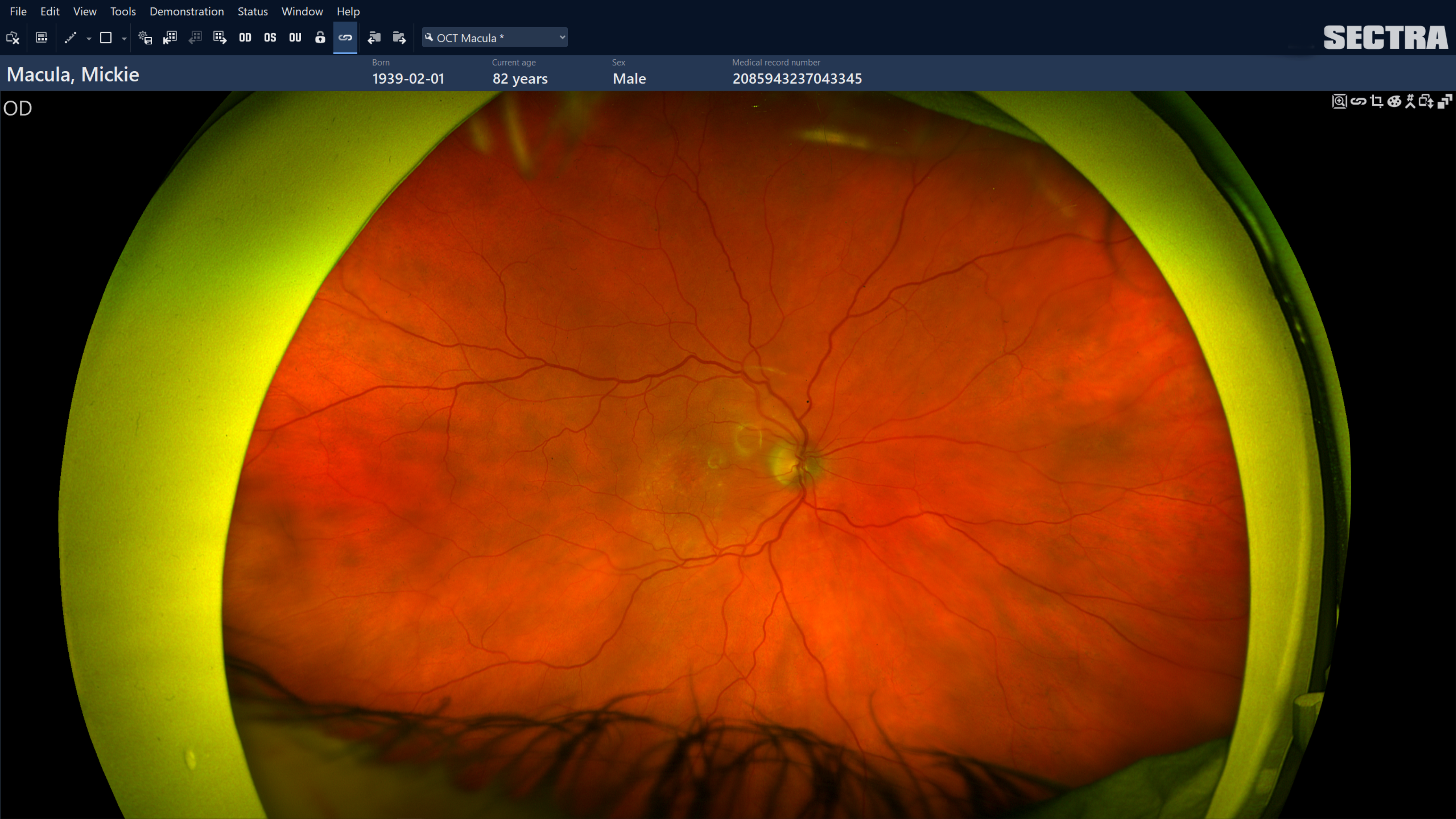
For medical imaging, the main standard in use is DICOM (Digital Imaging and Communications in Medicine). Here we find well developed specifications for the vast majority of imaging examinations performed in eye care. Healthcare organizations looking to acquire new equipment, are advised to demand that all relevant parts of this standard are fulfilled for all examinations performed with the device.
The American Academy of Ophthalmology as well as the National Eye Institute both promote the benefits of this concept.

