Article

Prepare your enterprise imaging for digital pathology: 6 areas of guidance for a successful solution evaluation

The ongoing digitization of pathology is a transformative change that offers new opportunities to increase efficiency, especially in cancer care, where over 90% of all tumors are confirmed by pathologists[1]. With this in mind, many healthcare organizations are beginning to use digital pathology technology for primary diagnostic work and not only research. As the DICOM standard for pathology becomes increasingly widespread, many health providers are now seeking ways to efficiently adopt digital pathology.

The most efficient way to digitize pathology in a manner aligned with your current imaging strategy is to incorporate it into the enterprise imaging (EI) solution, instead of creating yet another departmental IT system. When it comes to selecting digital pathology software, CIOs face a challenge since only a few EI solutions have proven (as of yet) to meet the tough performance demands of viewing and storing whole slide imaging (WSI) in a full clinical production environment.

This article will provide six areas of guidance in selecting and evaluating a solution that will allow you to reap the full benefits of digital pathology by adding this discipline to your EI solution.

Adding pathology to enterprise imaging—Why?

From an IT standpoint, the most obvious gains to be achieved by adding pathology to the EI solution, in contrast to digitizing pathology separately, is the ability to cut costs through the consolidation of IT systems and centralization of integrations. A standalone set-up will not be able to generate the same benefits in terms of clinical value, nor the cost advantages from an IT perspective, as an integrated multi-ology EI solution.

From a clinical point of view, having digital pathology as part of the EI solution will unite radiologists and pathologists, enhancing their collaboration through the use of a single diagnostic system. Initially, there are big wins to be gained simply by allowing different specialist groups to access each other’s images, test results and reports. This multidisciplinary convergence is encapsulated by the term integrated diagnostics (ID), the benefits of which are described in detail in an article[4] published in Radiology in 2017. The same article also highlights that now is the right time to unite pathology and radiology:

“This is the right time for a major move toward ID […] One major change is that pathology diagnostics is transforming from an analog-slide-and-microscope approach to a digital workflow at a rapid pace thanks to whole-slide imaging scanners and pathology picture archiving and communication systems (PACS) for large-scale, clinical use.”

One clinical benefit of using a single multi-ology solution achieved early on is increased efficiency in the preparation, presentation and follow-up of tumor boards. For early adopters of adding pathology to the EI solution, access to clinical information and increased efficiency in tumor boards have exceeded the expectations of specialists involved in cancer care. In a case study[5] at University Hospitals of Cleveland (OH), Dr. Hannah Gilmore, Division Chief of Anatomic Pathology and Director of the Breast Pathology Service, says:

“Since the pilot, we’ve been uploading pathology and radiology images into the enterprise PACS and projecting images from both departments on one system for each monthly conference. Using one system to display the images has streamlined how the cases are viewed, annotated and presented, and has greatly increased efficiency.”

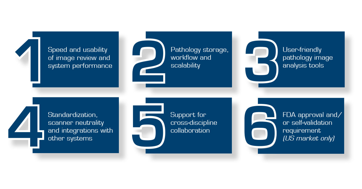
Six areas to look into when digitizing pathology

There are quite a few EI solutions on the market today claiming to be able to handle digital pathology. The differences between them were highlighted, for example, in the first pathology-specific report published by KLAS Research in January 2020[6].

But supporting pathology is about much more than just being able to store WSI. We have listed six areas, along with guidance on what requirements an EI solution needs to fulfill in order to provide a fully digital and microscope-free pathology workflow, capable of gaining pathologists’ acceptance, while also allowing clinical and cost benefits to be realized using a single EI solution. These are:

Six areas of guidance for a successful solution evaluation

1. Speed and usability of image review and system performance

The speed of the image viewer is at the very core of securing pathologists’ acceptance and getting them to let go of the microscope. It needs to be intuitive when it comes to image navigation, fast and not pixilate. Pathology images are large—sometimes several gigabytes in size—and to offer high viewing performance, modern digital pathology solutions use technologies such as web, data streaming and server-side rendering. Simply put, the EI solution must offer high speed in terms of viewing.

How the viewer is designed, and the way functionality is integrated into the user interface have also proven to be significant. An optimized and intuitive user interface is essential for minimizing the number of clicks and providing efficient tools for review. Information such as tissue registration data and details about the patient also needs to be presented to the pathologist in an easy-to-find and logical manner.

Ergonomics have historically been an important driver for digitizing pathology to avoid neck and shoulder problems caused by working with microscopes[7]. However, the computer mouse has turned out to be strenuous for the wrist when used continuously. Hence, it is necessary to evaluate the interaction devices with which the solution can be used. A study[8] comparing different interaction devices for navigating digital pathology slides suggested that a 3D mouse was the most appropriate option. The performance of the interaction device depends on both the device itself and how well its commands have been integrated with the viewer, meaning that both factors should be evaluated.

In an analog workflow, a microscope is always available. A digital solution thus needs to deliver extraordinarily high system availability and robustness. It should be capable of delivering the same performance regardless of whether there are one or a hundred users logged in looking at the same image, one or fifty slides per case, or whether a slide is 0.5 or 150 GB.

Overall, we see many EI vendors claiming they support digital pathology, but very few truly meet the performance requirements necessary to satisfy the pathologists’ demands.

2. Pathology storage, workflow and scalability

Digital pathology is a key component when it comes to reaping the full potential of enterprise healthcare systems, mergers and acquisitions. Digital referrals, reports, dynamic worklists and digital images enable both workload balancing and sub-specializations to be achieved. This is essential to realizing high efficiency and full utilization of experts to reach the best diagnostic quality across the enterprise.

A good digital solution should not only move the diagnostic review from the microscope to a monitor, but also offer tools to replace the previous workflow centered around glass trays and paper referrals. Necessary tools include customizable dynamic worklists as well as showing the required patient information from current and prior referrals, reports and images. Not to be forgotten is the fact that a digital pathology solution should also offer workflow support for the technicians that handle the quality assurance of the images right after the glass slides have been scanned.

Other tools to look for are those that have traditionally been offered in the Laboratory Information System (LIS), but where efficiency could be increased by instead moving these tools to the image-centric solution. Such tools include speech recognition and customizable structured reports, which can significantly enhance the reporting and follow-up workflow.

One benefit of digital pathology that is not offered by an analog workflow is the fact that users can access images at anytime from anywhere. First-generation viewers, such as those associated with a scanner, would normally not support this. Second-generation applications, which we refer to in this article as digital pathology solutions (as part of the EI solution), allow users to access the central EI archive from any computer within the network—without moving the large images around.

Using the EI solution for digital pathology also allows for centrally administered workflow orchestration, enabling the right case to be distributed to the right pathologist at the right time. This is something that will become increasingly important as pathology labs and health providers in general consolidate.

As the digitization of pathology often takes place in an iterative manner, the solution should be scalable and able to grow without forklift upgrades as the number of users and amount of data increase. In the early phases of digitization, the need for image storage may be relatively small, but as production increases, the benefits of functions often offered by the EI solution, such as image lifecycle management (ILM) and tiered storage, will be more obvious. ILM includes rules that can be applied to the image storage to steer which images to save and which to delete in order to reduce the cost of storage. Tiered storage optimizes costs by making sure you utilize fast disc space for the cases likely to be reviewed within a short period of time, and cheaper but slower disc space for long-term storage.

3. User-friendly pathology image analysis tools

Pathologists today face increased pressure as a result of steadily growing volumes and increasingly complex image reviews. The impending shortage of pathologists and ongoing prevalence of burnout will further strain healthcare over the next few years and onward. A survey[9] (15,000 respondents in October 2018) published in the Medscape National Physician Burnout, Depression & Suicide Report 2019 found that an incredible 33% of US pathologists have reported burnout symptoms during the last year. Two of the most common factors leading to burnout are having too many administrative and time-consuming tasks and spending too many hours at work[10].

Digital pathology offers functionality beyond the microscope that prevents burnout by providing opportunities to increase efficiency in diagnostics. Previously manual tasks can be facilitated or automated by using user-friendly image analysis tools that can assist, for example, in counting cells, performing percentage cancer tissue calculations or other time-consuming tasks.

Another advantage of utilizing image analysis algorithms is that they can improve quality and standardize the review to make it more consistent between pathologists. Using the same quantification tools rather than manual quantification can lead to less variability, enabling pathologists to deliver more consistent results[11]. For most pathology departments, the realization of the efficiency and quality gains that come with image analysis is a prerequisite to justify the additional step of scanning which digital pathology involves.

Traditional glass review offers pathologists a variety of ways to customize the microscope to fit individual preferences. In a similar way, pathologists’ preferences when it comes to algorithms vary. Moreover, the technology and the vendors behind the image analysis and AI applications evolve very quickly, with new vendors entering, being acquired or leaving the market. These factors combined reinforce the importance of the EI solution’s ability to offer a full portfolio of image analysis applications from many different vendors. In addition to a broad portfolio of applications, the importance of a tight integration with the viewer and workflow, together with high usability, cannot be emphasized enough.

4. Standardization, scanner neutrality and integration with other systems

“Pathology-enabling” your EI solution means that additional types of modalities, i.e. scanners, need to be connected. Just as in the early days of digital radiology, pathology images today are often produced in proprietary file formats depending on the scanner vendor. However, a shift towards a common DICOM standard is becoming more widespread, with most of the big scanner vendors now able to produce images in a DICOM for WSI format.

Using pathology images in DICOM format offers many benefits, primarily since it clearly separates the image capture devices (the scanners) from the viewing and workflow solution (the EI solution). In an environment where DICOM is fully implemented, this will provide the freedom to use any scanner and view images from anywhere. Since the DICOM standard for pathology is not yet as widely adopted as it is in radiology, it is important to make sure that the EI vendor has plans to fully support the standard when it comes to storage, viewing and communication.

To reap the potential benefits from the start and in the future, it is important that your EI solution supports DICOM according to Supplement 145, as well as the most common proprietary file formats. Supporting proprietary file formats enables collaborations with other hospitals that do not produce images according to the DICOM standard.

For cost-effective integrations with the rest of the surrounding healthcare IT environment, the EI solution needs to support the available standardized integration protocols, such as HL7 and FHIR. There is plenty of experience from other integration projects in healthcare, including EMRs, PACS and RIS, to confirm the value of standardized integrations. By the same token, the digital pathology part of the EI solution should comply with HL7 in order to facilitate a seamless integration with the LIS and the EMR, which is crucial from a pathology workflow perspective.

Another aspect worth considering is to look at the EI vendor’s proven track record of integrating with EMR vendors. An EI solution consists of many different modules with high demands in terms of being kept up to date, and for each new release the vendor must ensure the integration remains whole. The vendor’s capability to keep integrations stable and collaborate with EMR vendors is key.

5. Support for cross-discipline collaboration

Key specialists in cancer care, including pathologists, radiologists, oncologists and surgeons, have traditionally worked in separate, departmental IT silos, hindering them from sharing images, test results and reports with each other in a joint system. With the introduction of enterprise imaging, this is changing, enabling more efficient cross-discipline collaboration. The digitization of pathology is the last remaining piece of the puzzle.

In addition to gaining access to other specialists’ images and reports, one of the key benefits of using an EI solution that supports digital pathology is that it facilitates the preparation, presentation and follow-up of tumor boards. These meetings are very expensive, and functionality that can facilitate associated tasks will result in significant cost savings by reducing the time spent per patient.

To save time, an EI solution should offer tools that allow pathologists to add cases to a specific tumor board in conjunction with the diagnostic review instead of afterwards, as is the case in an analog workflow. It should also be able to present images and case information from radiology, pathology, oncology, surgery and other specialties during the meeting itself from a single user interface, simply by accessing the right worklist. Evidence has shown that efficient tumor boards improve diagnostic accuracy, adherence to clinical practice guidelines and some clinical outcomes[12]. Hence, tools that can facilitate tumor boards will have an impact both clinically and in terms of costs.

Using one system to display the images has streamlined how the cases are viewed, annotated and presented, and has greatly increased efficiency. […] Most importantly, we can make sure that the patient is getting the most appropriate treatment. More access, better access and shorter wait times will help too. [13]

Hannah Gilmore, MD, Division Chief of Anatomic Pathology and Director of the Breast Pathology Service at University Hospitals of Cleveland, OH

6. FDA approval and/or self-validation requirement (US market only)

Selecting an EI vendor that supports digital pathology is important, but you should also be aware that the US Food and Drug Administration (FDA) requires an approval or 510(k) clearance for vendors to market and sell solutions for digital pathology primary diagnostic review. A US health provider that wishes to use an EI solution for primary diagnostics in pathology is not obligated to use an FDA-approved solution, but rather can choose to perform a self-validation of a non-approved system.

At the time of writing (January 2020), there are two approved/cleared digital pathology systems, while several more are pending approval or are in the process of submitting their application.

Note: Vendors without US FDA approval are allowed to sell and market their digital pathology solutions for tumor boards, frozen sections and consultations.

Summary

The digitization of pathology is here and involves investments in new IT systems for image management and diagnostics. If introduced wisely as part of the enterprise imaging solution, it will provide major opportunities to improve the efficiency and quality of cancer diagnosis. From an IT perspective, this also means that the existing enterprise platform can be further utilized, economies of scale realized, and new departmental IT silos avoided.

Many enterprise imaging solution vendors claim they can handle digital pathology, but it takes more than simply introducing a system that can store WSI to support a clinical pathology workflow and replace microscopes. The digitization of pathology is a transformative change that places stringent requirements on the entire enterprise imaging solution to support such a broad-scale transition. This article has provided six areas of guidance to make sure you select a digital pathology solution as a part of your enterprise imaging solution that can successfully help your organization to reap the benefits of the coming transformation.

References

  1. https://training.seer.cancer.gov/casefinding/sources/pathology.html
  2. https://jamanetwork.com/journals/jamanetworkopen/fullarticle/2734800
  3. https://klasresearch.com/report/global-digital-pathology-2019/1349
  4. https://pubs.rsna.org/doi/full/10.1148/radiol.2017170062
  5. https://medical.sectra.com/case/digital-pathology-pilot-predicts-prosperity/
  6. https://klasresearch.com/report/global-digital-pathology-2019/1349
  7. http://www.jpathinformatics.org/article.asp?issn=2153-3539;year=2014;volume=5;issue=1;spage=14;epage=14;aulast=Thorstenson;type=0
  8. J. Molin, C. Lundström, M. Fjeld, 2014, A comparative study of input devices for digital slide navigation, J Pathol Inform 2015;6:7.
  9. https://www.medscape.com/slideshow/2019-lifestyle-burnout-depression-6011056#3
  10. https://www.medscape.com/slideshow/2019-lifestyle-pathologist-6011146#6
  11. Stålhammar, Gustav, et al. “Digital image analysis outperforms manual biomarker assessment in breast cancer.” Modern Pathology 29.4 (2016): 318.
  12. https://www.ncbi.nlm.nih.gov/pubmed/23891067
  13. https://medical.sectra.com/case/digital-pathology-pilot-predicts-prosperity/
Author: Simon Häger, Market Strategist at Sectra

Related reading

Related products