Exploring anatomy in 2D, 3D, and cross-sections
Develop a deeper understanding of anatomy, spatial relationships, and pathology through real clinical imaging.
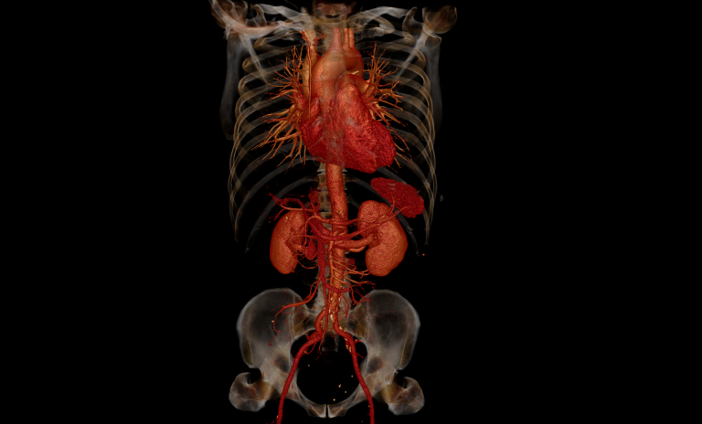
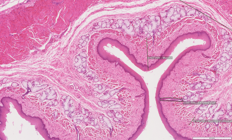
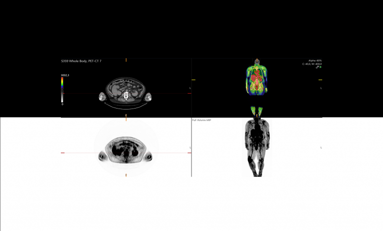
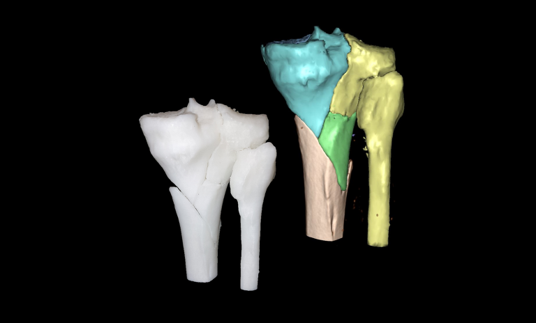
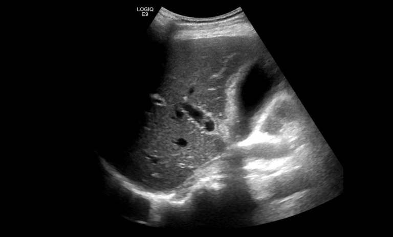
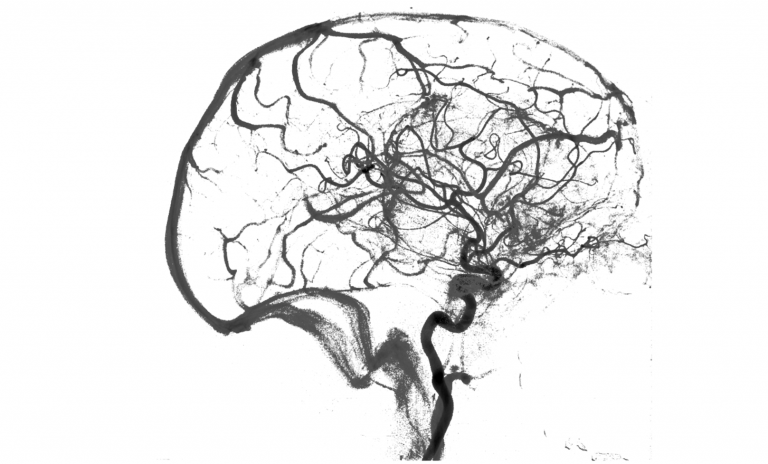
The Sectra Education Portal allows users to explore normal and pathological anatomy using different imaging modalities, including 2D (multiplanar views) and 3D reconstructions. It also includes an interactive anatomy atlas and visualization tools, allowing users to compare cross-sectional images, surface anatomy, and histological structures—helping bridge the gap between imaging and clinical reality.