Region Dalarna

Integrated diagnostics in practice: How Region Dalarna united radiology and pathology for improved breast cancer care

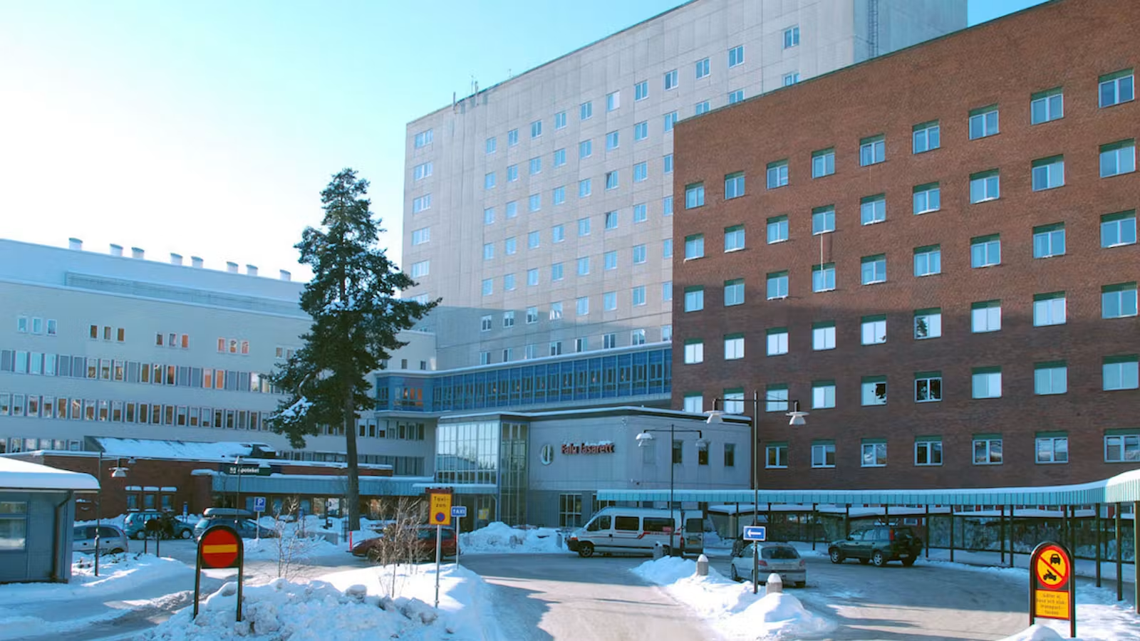
Region Dalarna, a Swedish healthcare region serving around 290,000 residents, with its main hospitals in Falun and Mora, has brought digital pathology into the same Sectra platform already used by its radiology and mammography departments. The integration means that radiologists and pathologists now work from a shared diagnostic environment, accessing each other’s images and findings without switching systems. To understand what that means in practice, we met Helena Cederberg, manager of digital pathology and system administration at the laboratory, and Annette Bauer, a pathologist specializing in breast pathology. We also spoke with the mammography team: Cathrin Döbereiner, mammographer; Anita Johansson, site manager; and Elin Svartling, medical secretary and system administrator.

The change for Region Dalarna is most visible in the workflow surrounding breast cancer surgery. When a surgical specimen arrives, it travels from theatre to mammography for specimen radiography, then to the pathology lab. Helena Cederberg describes how, at each step, the previous step’s findings are now immediately visible.

“The pathologists can see what descriptions the radiologists have made,” she explains. “If the radiologists want it sectioned in a certain way, we do that. And it gives us guidance on how to cut, especially for our large sections.”

Even for small tissue fragments being examined for calcifications, the digital image provides orientation, a hint about where in the tissue to focus attention. For the lab, it removes the guesswork from a step that directly affects what gets examined.

We have become more efficient, and it has improved the quality.

Annette Bauer, Pathologist, Region Dalarna

Annette Bauer has worked with Sectra since before the department went fully digital. When digital pathology was integrated into the platform, histological slides joined the radiology images in a single shared environment, giving pathologists and radiologists access to each other’s findings.

That shift matters most in complex cases, such as tumors with diffuse growth patterns, multifocal disease, or situations where understanding the spatial distribution of a lesion is critical for surgical planning. With both imaging and tissue findings in the same session, Bauer can correlate them directly, rather than reconstructing the relationship from memory or separate screens.

“We can more easily correlate histological images with specimen radiography,” she says. “The distribution of the lesion we are examining—we can correlate that very well. And especially when you have them in the same system.” Previously, this kind of comparison meant projecting a microscope image onto a screen and comparing it manually. Now, both can be displayed and navigated side by side. “We have become more efficient, and it has improved the quality,” she adds.

That improvement in efficiency and quality is reflected in how cases are reviewed and discussed across the breast cancer team. For the mammography team, the most tangible benefit shows up in preparation for postoperative meetings and multidisciplinary team meetings. Previously, pathological images were only visible during the meeting itself. Cathrin Döbereiner describes the difference:

“Before, we saw these images at the multidisciplinary team meetings,” she says. “Now we can, in peace and quiet, compare and check—what we said, what we saw. You can prepare yourself in a much better way.”
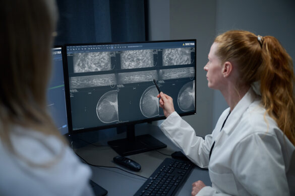
Being able to show and navigate digital histological images during discussion—­­­­pointing to margins, demonstrating tumor distribution—changes what can be communicated.

“It is easier to show things on a digital histological image”, says Bauer. “It helps us speak the same language, to see the same thing.” For a department where radiologists and pathologists now share a single system, that shared view has become the foundation for how cancer cases are discussed. “It is always a good solution to be in the same system,” she says. “It facilitates communication.”

The pathology department has long used large-format tissue sections, roughly twice the size of a standard mega sections, sometimes referred to as mega slides, to examine the full breast specimen surface in a single preparation. Digitalization has extended the value of this technique­—several sections can now be displayed side by side, annotated, and compared on screen.

“A larger slide gives me the possibility to examine a larger tissue surface at once,” says Bauer, “and I get a better understanding of the tumor and its surroundings.” For cases with complex tumor architecture or multifocality, this overview changes what is visible and what can be communicated to the surgeon.

Ultimately, this shared understanding between radiologists and pathologists supports better-informed treatment decisions for patients. The specialists involved in their care can review the same information, discuss findings more thoroughly, and make decisions based on a more complete picture of the disease.

It is always a good solution to be in the same system. It facilitates communication.

Annette Bauer, Pathologist, Region Dalarna.

Beyond the direct diagnostic workflow, the shared platform has enabled a more flexible work environment. Pathologists can now work remotely and internal consultations that previously required shipping glass slides now happens within the system.

“It has made remote work and internal consultations possible”, says Bauer. “Our locum pathologists can work from home with a company computer. That was not possible before.”

When looking ahead, one feature the mammography team sees as an untapped opportunity is the usage of the chat function within the platform between the mammography team and the pathologists. Elin Svartling, medical secretary and system admin at the mammography department, notes that the technical capability to communicate directly with pathologists within the platform already exists, it simply has not yet become part of the routine.

“It is something that could be improved, that you send a patient case and talk directly”, she says. For a team that already shares a platform, it feels like a natural next step, she means.

For Bauer, the integrated platform is also the foundation for what comes next. Looking ahead, she sees significant potential in AI-assisted image analysis, including algorithms designed to support prognostic assessment in breast cancer. While the department is still in the process of evaluating and implementing these tools, she believes they will become an increasingly important complement to both traditional morphology and molecular pathology. Building on the collaborative workflow that already brings radiology and pathology together in a shared environment, she sees AI as another step toward more informed diagnostic decisions and more personalized patient care.