Annette Bauer has worked with Sectra since before the department went fully digital. When digital pathology was integrated into the platform, histological slides joined the radiology images in a single shared environment, giving pathologists and radiologists access to each other’s findings.
That shift matters most in complex cases, such as tumors with diffuse growth patterns, multifocal disease, or situations where understanding the spatial distribution of a lesion is critical for surgical planning. With both imaging and tissue findings in the same session, Bauer can correlate them directly, rather than reconstructing the relationship from memory or separate screens.
“We can more easily correlate histological images with specimen radiography,” she says. “The distribution of the lesion we are examining—we can correlate that very well. And especially when you have them in the same system.” Previously, this kind of comparison meant projecting a microscope image onto a screen and comparing it manually. Now, both can be displayed and navigated side by side. “We have become more efficient, and it has improved the quality,” she adds.
That improvement in efficiency and quality is reflected in how cases are reviewed and discussed across the breast cancer team. For the mammography team, the most tangible benefit shows up in preparation for postoperative meetings and multidisciplinary team meetings. Previously, pathological images were only visible during the meeting itself. Cathrin Döbereiner describes the difference:
“Before, we saw these images at the multidisciplinary team meetings,” she says. “Now we can, in peace and quiet, compare and check—what we said, what we saw. You can prepare yourself in a much better way.”
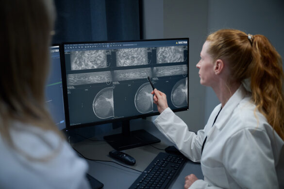
Being able to show and navigate digital histological images during discussion—pointing to margins, demonstrating tumor distribution—changes what can be communicated.
“It is easier to show things on a digital histological image”, says Bauer. “It helps us speak the same language, to see the same thing.” For a department where radiologists and pathologists now share a single system, that shared view has become the foundation for how cancer cases are discussed. “It is always a good solution to be in the same system,” she says. “It facilitates communication.”
The pathology department has long used large-format tissue sections, roughly twice the size of a standard mega sections, sometimes referred to as mega slides, to examine the full breast specimen surface in a single preparation. Digitalization has extended the value of this technique—several sections can now be displayed side by side, annotated, and compared on screen.
“A larger slide gives me the possibility to examine a larger tissue surface at once,” says Bauer, “and I get a better understanding of the tumor and its surroundings.” For cases with complex tumor architecture or multifocality, this overview changes what is visible and what can be communicated to the surgeon.
Ultimately, this shared understanding between radiologists and pathologists supports better-informed treatment decisions for patients. The specialists involved in their care can review the same information, discuss findings more thoroughly, and make decisions based on a more complete picture of the disease.