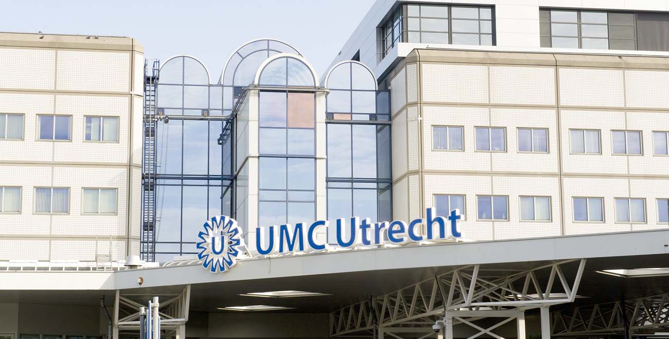
At the beginning of 2016, University Hospital Utrecht (UMCU) became the first hospital in the Netherlands to digitize its entire pathology workflow. What has happened since? And how are they utilizing digital pathology to move forward within integrated diagnostics and machine learning?
It is more efficient, and the risk of errors is smaller, meaning patient safety increases. I can’t really come up with a better argument for switching to digital pathology.
For Prof. Dr. Paul van Diest, head of the pathology department at UMCU, it has been clear for a long time that digital image analysis applications based on machine learning provide countless possibilities for pathology. When it became possible to quickly create a high-resolution digital image of a glass slide, he didn’t want to wait until the technology was fully developed. Instead he took the lead himself.
Following a tender process, UMCU selected Sectra’s digital pathology solution. Together they developed on the foundation that Sectra had already designed. Van Diest involved the entire pathology department. “Not everyone was enthusiastic about digitizing from the start. First, you must gain trust and build up experience. Sectra has extensive experience of digitizing radiology. The paths of radiology and pathology are very similar, and Sectra was therefore able to guide us very well through the process of going digital—they knew what we would encounter.”
Working groups designed the digital process
To gain trust and support, almost everyone in the pathology department was assigned to a working group during the development phase. Each group focused on a specific part of the process. The working groups had full mandate to solve the challenges they were given. “That was quite exciting,” says van Diest. “As a department head, I suddenly had nothing left to say. The working groups were in charge. That led to a huge involvement in the workplace.”
Sectra’s solution went live in early 2016. A year and a half later, it is estimated that more than 80 percent of the diagnostics are fully digital. Van Diest comments, “We will never grow to 100 percent, because some reviews can’t be done digitally, for example, cases that require birefringence. However, I would be very happy if we ultimately reach 95 percent.”
Prof. Dr. Marijke van Dijk has already reached 95 percent. She specializes in skin review. “I found it quite exciting because I have the largest production in the entire department. You can do my type of review quickly. However, before going digital, a lot of time was spent matching the different elements needed. With digital pathology, you always have the patient information, images, speech recognition system and report together automatically. I now waste less time matching these.”
A digital display also works very well for her type of review, she says. “I look at patterns and that works very well on a screen. Skin samples are generally small and can easily be captured in one image. I can zoom in as much as I want.”
The first few weeks, van Dijk performed double reviews, but soon she said goodbye to the microscope for most of the diagnostics. “There are two types of review in which a digital image is less accurate, for which I always use the microscope. I estimate that this represents at most 2 percent of all glass slides.”
Van Diest says he works almost 100 percent digitally. “That is because the area I am specialized in, breast abnormalities, suits digital review very well. I almost never have to fall back on a physical glass slide.”
Digital work also offers many advantages in the field of nephropathology. According to Dr. Tri Nguyen, who specializes in kidney abnormalities, the most important thing is that digital images have an unlimited shelf life. “I regularly work with review methods where the staining fades over time. Then there is no point in saving a glass in an archive. This is annoying if a patient gets ill a few years later, because then you can only search for the report but not the section itself. And it is also unfortunate because the insights in our field continue to change. For example, we regularly discover new diseases. It would be interesting to find out now, in cases where we couldn’t make a good diagnosis ten years ago, whether it was a disease that we just discovered. And I’m not even talking about the chances of being able to gain new insights with the help of big data analytics. If you have a digital image archive, you can efficiently search for patterns and connections. This is extremely difficult based on reports alone.”
We still work with separate systems next to each other. The next step is to make all medical image information accessible via a single entry […] This way we can grow towards more integrated diagnostics.
Integrating radiology and pathology
UMCU is working together with Sectra to improve and extend the existing solution. There is currently a list of dozens of ideas for improved and new functionalities that can be developed by Sectra, and the development priorities are determined periodically in consultation with a team in Sweden.
High up on the agenda is integrating radiology and pathology into a single system, so that integrated diagnostics can be better supported. Van Diest explains, “Here at UMCU, we still work with separate systems next to each other. The next step is to make all medical image information accessible via a single entry, so that you don’t have to log into different systems all the time and making it easier to access a radiology image or other essential information. This way we can grow towards more integrated diagnostics. Of course, everyone keeps their own specialization, but we can break down the walls that still exist to some extent.”
Van Dijk fully agrees with him. “We never used to look at glass slides during oncology discussions. It sometimes happened that a plastic surgeon wanted to see a slide. Then he had to go to my department and look through a microscope. Now I can show the digital image during a meeting. I notice that this sparks interest in my field. As a result, I’m more often asked to look at an image after a discussion. And that can be done more easily now, because you do not need a microscope for that. It is much more pleasant to sit together in front of a screen.”
We hope that the things we discover and the things they develop will reinforce each other—that we can build on each other’s insights.
Machine learning in pathology
As mentioned, van Diest is already experimenting with machine learning. “We are already able to develop an algorithm with less than one pathologist in two weeks’ time. That is great support for our work. The challenge, however, lies not so much in the development of algorithms, but especially in the integration into the daily workflow.”
Sectra offers a vendor-neutral interface to such algorithms and applications in its platform. This means that customers can utilize their own machine learning applications as well as Sectra’s and virtually any other application—regardless of vendor—through a single, unified workplace. Sectra’s own efforts within machine learning mainly focus on applications for improving the clinical workflow and increasing the productivity of radiologists and pathologists. Development is carried out both in-house and in collaborative settings.
Van Diest and Nguyen are happy that Sectra is also working on machine learning. “We hope that the things we discover and the things they develop will reinforce each other—that we can build on each other’s insights.” Such a co-creation-oriented relationship is also exactly what Sectra is looking for. Sectra needs the input of passionate pathologists, such as van Diest, Nguyen and van Dijk, to bring new technology in line with real needs that exist in the market.
Create support for digital work
If the pathologists at UMCU were to give one piece of advice to other hospitals, it would be: reap the benefits of digital work.
Tri Nguyen concludes, “It will take a long time before the quality of the digital images is as high as that of the physical glass in all types of review. Do not wait for that. It is not ‘all or nothing’. Initially, you will still have to keep a physical and digital workflow in parallel for a period, not least because pathologists must be given the time to learn to trust digital sections. The longer you work digitally, the less often you take a physical glass. In our lab, we now work primarily digitally, unless the type of review doesn’t allow it. Simply because it offers so many benefits. It is more efficient, and the risk of errors is smaller, meaning patient safety increases. I can’t really come up with a better argument for switching to digital pathology.”
Featured products & services
Related cases


