Region Värmland

Swedish county council shows the way to a successful implementation of enterprise imaging

Region Värmland is a pioneer in enterprise imaging and is currently a world leader in managing and storing medical images, multimedia and patient information in a central, region-wide platform. In a previous article, we described how the initial introduction went in 2014, when the county council expanded its radiology PACS to an enterprise imaging system. How has the continued implementation helped connect additional fields, such as anatomical pathology, endoscopy and speech-language pathology? Which clinical and organizational benefits have they experienced so far? And what do their future plans look like for managing patient-generated images and engaging patients more in their own care—a topic of broad and current interest in Swedish healthcare? We met with departmental representatives who described the path to success in their own words.

Always having access to the most relevant and comprehensive information on which to base our decisions has improved both the efficiency and quality of our diagnostics and follow-up treatment.

Marie Moberg, PACS/VNA System Administrator for Region Värmland

The shared image archive improves the flow of care. Images can now be taken directly at the primary care center and analyzed by specialists as needed. This means we can treat the patient correctly and avoid unnecessary extra visits—which is good for both patients and healthcare providers.

Thomas Miliander, Head of IT and Business Support for Region Värmland

From digitization in radiology to comprehensive enterprise imaging

Region Värmland is taking steps to realize the vision that started the entire project back in 2012: to implement “a shared archive for ALL departments and ALL file types.”

“When we decided to implement a department-wide multimedia system, we chose to expand our existing radiology PACS from Sectra,” says Thomas Miliander, Head of IT and Business Support. “PACS supports the prevailing standard in medical imaging, DICOM, and the system is also scalable and designed to manage large volumes of images and many users.”

Focus on connecting more fields and disciplines

In 2012, new functionality was installed in the PACS so that the journey towards a comprehensive enterprise imaging system could begin. One after another, fields outside radiology were connected to the system, which gave specialist clinics, departments and district health centers the ability to download images, videos, sound files, documents and reports directly from the platform. Thomas explains that as much information as possible is saved in DICOM, but that since this is not possible for certain types of multimedia that are not yet supported by the standard, they are saved in other formats such as PDF, WAV, etc. The types of multimedia currently handled within the system include:

  • Radiology examinations
  • Digital pathology images
  • Skin images
  • Vision tests and images from eye clinics
  • Surgery videos
  • Endoscopy images
  • Sound files from speech pathologists
  • Ultrasounds from the emergency room, ICU, surgery, pediatric clinics, cardiologists, women’s clinics and others
  • Colonoscopy images from women’s clinics
  • Spirometry reports and broncoscopy images from pulmonary clinics
  • Bone density measurements from endocrinologists and diabetes centers
  • Several types of other “-scopy” images (in addition to those named above): cystoscopy, stroboscopy, laparoscopy

Important steps taken in 2018

Marie Moberg is the System Administrator for Region Värmland’s PACS/VNA and has been part of the journey since the end of 2013. She describes 2018 as an exciting year when several important activities were conducted and real progress was made towards implementing their vision. Among these, she highlights the following cornerstones:

  • The first stage in digital pathology was completed and all the minor cases in histopathology were scanned and saved digitally.
  • A system for video management (recording, displaying and editing) was introduced, including a mobile app for capturing images and videos.
  • The universal viewer was made available to all departments and from all types of devices.
  • An XDS archive was set up to save all file types.
  • It was ensured that the platform and the viewer fulfill all legal and security requirements and that the archive contains high-quality information.

In addition, over 150 clinics and healthcare departments were connected during the year, allowing them to store and consume multimedia through the platform every day. “As the system was expanded to more departments, several others also noticed the gains that could be achieved, which meant that the solution developed a good reputation within the county council and even more areas decided to join,” continues Marie. The platform developments that took place in 2018 put Värmland on the map as a world leader in enterprise imaging. Marie and Thomas reveal that the progress made has attracted considerable outside interest and that they have already hosted a number of visits from other county councils and healthcare systems—not just from Sweden, but from several countries around the world.

These days, I save five to ten minutes per patient for every ultrasound examination.

Edit Floderer, Senior Physician and Cardiologist at the Cardiology and Intensive Care Unit at Central Hospital of Karlstad

We expect major gains in the future as a result of the digitization of pathology, including being able to share cases digitally instead of sending slides, being able to apply automated image analysis and improving the quality and efficiency of the workflow when the handling of slides can be eliminated after they’re scanned.

Charlotta Gestblom, Head of the Pathology Department at Central Hospital of Karlstad

Benefits of enterprise imaging

Patients are often transferred between different departments that produce images, videos, sound files and documents so that specialists can make the correct diagnosis and provide the right treatment. The key argument for patient-centered care is that all of this information should follow the patient and be made available to the relevant parties regardless of where the patient is in the healthcare process. This is the primary problem that an enterprise imaging system is designed to solve—something that is very clear to the system’s users in Region Värmland.

“In general, increased access to multimedia and patient information is highlighted as the absolute most important advantage in the organization,” says Marie. “No longer needing to spend time searching and always having access to the most relevant and comprehensive information on which to base our decisions has improved both the efficiency and quality of our diagnostics and follow-up treatment.”

Marie further explains that many specialists say they were previously unable to use the patient’s complete diagnostic history since it was hard to access. By storing everything in the same solution, all relevant examinations can more easily be taken into consideration, and it even becomes easier to compare different examinations over time in order to measure the progress of a patient’s treatment. This not only provides major clinical value, it is also valuable from a pedagogical perspective in conversations with patients.

Multidisciplinary rounds have also become much more effective since all relevant multimedia can be reached from a single system. Documentation from radiology, pathology, dermatology and oncology are collected in one place and can be retrieved with a viewer, which means that specialists can easily prepare and present material, and see their colleagues’ answers and conclusions beforehand. This way, everyone is better prepared during the rounds, which means they can discuss more patients in the same amount of time.

Thomas highlights additional efficiency improvements with the enterprise imaging system. “The shared image archive improves the flow of care. Images can now be taken directly at the primary care center and analyzed by specialists as needed. This means we can treat the patient correctly and avoid unnecessary extra visits—which is good for both patients and healthcare providers.”

Probably one of the most important advantages will be in the roll-out of AI applications, since these require access to a large amount of centralized and aggregated data. “By building an archive of different types of images and patient information, we are creating a solid foundation in order to quickly leverage the benefits of AI when new applications are released,” says Thomas.

He continues: “Overall, the introduction of the platform has also helped us reduce the number of IT systems, which provides cost benefits in terms of reduced maintenance, integration and avoiding different suppliers blaming each other if problems occur.”

Case: Cardiology

A clear example of immediate efficiency gains is highlighted by Edit Floderer, Senior Physician and Cardiologist at the Cardiology and Intensive Care Unit at Central Hospital of Karlstad, who explains that she currently saves five to ten minutes per patient for every ultrasound examination.

“Connecting the ultrasound apparatus directly to the enterprise imaging system via Wi-Fi saves time both at the beginning and end of every examination. Bookings can be made quickly in the EMR ahead of time, and when it’s time for the examination all we have to do is choose the patient in the list on the instrument,” says Edit.

“After the examination, we just hit ‘save’ and the results then become directly accessible in the patient journal. The whole process used to be pretty disorganized, measuring the patient manually and then saving and transferring images,” she concludes.

Reducing the number of manual steps in the workflow saves several hours per week.

Case: Digital pathology

Sectra’s enterprise imaging system is based on the same technical platform for all image-intensive departments, including anatomical pathology, which is a major advantage in terms of collaboration and achieving an integrated workflow between the “-ologies.” In addition, Sectra has one of the only solutions on the market that also supports an efficient workflow and high-performance image viewing for digital pathology.

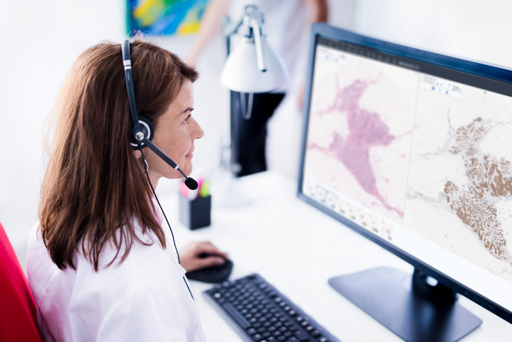
The Pathology Department at Central Hospital of Karlstad has scanned all of its minor histopathology cases and conducted a validation project to ensure that the diagnoses made using digital pathology images are of the same quality as those reviewed via microscope. The project is now continuing, with selected cases (prostate and liver) being scanned to enable primary reviewing.

“We expect major gains in the future as a result of the digitization of pathology, including being able to share cases digitally instead of sending slides, being able to apply automated image analysis and improving the quality and efficiency of the workflow when the handling of slides can be eliminated after they’re scanned,” says Charlotta Gestblom, Head of the Department of Pathology at Central Hospital of Karlstad. “Another major advantage is that we can more easily conduct rounds, both within the county of Värmland itself, but also at cross-regional and national levels.”

Another advantage that Charlotta expects to see in the future is the possibility for increased cooperation with radiology during rounds. “By using the same system for both diagnostics and image storage, we can access each other’s images. This is also an advantage when it comes to cooperating with other specialists—for example, accessing images from the dermatology clinic before diagnosing changes in skin conditions.”

It feels like I’m working in a single system.

Jenny Gustavsson, Speech-Language Pathologist at the Speech-Language Pathology Unit at Central Hospital of Karlstad

Basing our work on the users’ needs has been a major factor in our success.

Mikael Borén, CIO for Region Värmland

Success factors

Region Värmland has clearly succeeded in consolidating its image and multimedia management. But which factors do those involved believe played an important role in this success?

Mikael Borén, the county council’s CIO, says that tight integration with surrounding IT systems has been a key factor in creating a well-functioning workflow. This included integration with the EMR, lab systems and several specialist-specific diagnostic systems. “Because the EMR controls a large portion of the hospital’s workflow, close integration with this system made it easier to roll out the enterprise imaging platform, since the users perceive it as a single system,” says Mikael.

Mikael also emphasizes that it is incredibly important to have the right expertise in place from the beginning. “Basing our work on the users’ needs has been a major factor in our success. This hasn’t been just an IT project—representatives from the clinical workflow have also been involved, and we’ve really tried to listen and adapt to their actual needs.”

Marie, the PACS/VNA System Administrator, explains the importance of starting right away, on a small scale, and not planning too far ahead. “Begin with an introduction, learn, adjust and keep going. You can fine-tune functionality, flow and configuration after the fact. The project team has focused on informing users about the possibilities the platform offers instead of forcing it on them. This means that pioneers within the departments drove the implementation forward themselves. One clear example is Hans-Ulrik Stark, Chief Physician at the dermatology clinic. These days, he’s the one enthusiastically calling us for meetings so we can take the next step,” says Marie, smiling.

A healthy supplier collaboration has been essential to how smoothly the introduction has gone, according to Mikael. “We chose Sectra because our earlier collaboration had worked so well, and we realized that Sectra’s strategic focus was in line with our visions. The obvious dedication of Sectra’s employees also played a part in our selection. Product-wise, it was important for us to choose a supplier who had a stable system that supported standards and could integrate well with surrounding IT systems.”

Another important component that Mikael emphasizes was the frequent workshops that Region Värmland held together with Sectra and the county council’s EMR supplier.

Obviously, the implementation process was not entirely problem-free. Marie explains that one of the biggest challenges with implementing enterprise imaging was allocating user rights, since several types of users have access to the same system but all have different needs. “We solved that by giving users more general rights and a higher minimum license,” explains Marie. “So it was a matter of finding a balance between giving sufficiently broad access and limiting access to information that the user doesn’t need.”

Communicating internally about the possibilities that had been created also posed a challenge. “It’s been difficult to reach out to the hundreds of users, specialist clinics and primary care and outpatient facilities with information about how they can take advantage of the solution and start using it. The implementation could have gone faster if communication had been better. And for those who are already on board, we want to provide support in the form of training and further development in 2019—there’s a lot to build on,” Marie points out.

By building an archive of different types of images and patient information, we are creating a solid foundation in order to quickly leverage the benefits of AI when new applications are released.

Thomas Miliander, Head of IT and Business Support for Region Värmland

Expanded roll-out and visions for the future

Additional disciplines and departments are now ready to install the platform for their multimedia. “It’s not the technology that is limiting us. As I said, it’s about getting the information out about the possibilities available,” says Marie. She also explains that many departments are often pleasantly surprised at being able to use the platform, and that most of them are positive towards starting to use it.

Several pilot projects are currently ongoing—for example, in speech-language pathology, where sound clips are saved and can be accessed directly through the EMR. In pathology, discussions have begun concerning image lifetime management, which will be necessary when cytology is digitized due to the large amount of data. At that point, certain functions will need to be applied to either erase clinically irrelevant data or compress the examination. In the not-too-distant future, the plan is for molecular biology to start storing patient data in the platform as well.

Patient-generated data

A particularly exciting area is meeting the increased demand for utilizing patient-generated data, an area where Region Värmland has several activities under way. Many of the hospital’s departments have requested a function to allow patients to save images that they have taken themselves with their phones, something that will be possible in the near future.

“The challenge with saving patient-generated data isn’t about the technology, but about guaranteeing its quality,” says Hans-Ulrik Stark, Chief Physician at the Dermatology Department at Central Hospital of Karlstad. “We’ll most likely let a specialist be a ‘gate-keeper’ and approve the image before it’s saved in the system.”

Mobile devices

Using mobile devices, primary care and outpatient facilities can handle a larger number of cases, taking pressure off specialists and reducing the number of unnecessary patient visits. For example, images and videos can be recorded using an app in a work phone, and tablets can be used to look at multimedia with the universal viewer. The application of mobile devices also makes it possible for specialists to use different apps to support their workflow—for example, when saving questionnaires, documents and multimedia in the EMR and making them accessible.

With the ongoing introduction of touchscreens in the healthcare sector, specialists are no longer tied to a specific workplace, but can instead provide diagnoses and opinions in a more flexible manner. Mobile devices have also proven valuable from a pedagogical perspective when explaining things to and engaging patients.

Patient access to multimedia

Something that Region Värmland noticed early on was that patients increasingly want access to their medical images. Plans are in place to offer this service in a portal that will be integrated with the national healthcare e-service 1177 Vårdkontakter. Various portals are still being evaluated, including Sectra’s Image Exchange Portal (IEP), to determine which one will be used.

Giving patients access to their examinations would offer major benefits to individuals in the form of increased involvement in their own care and the ability to bring clinical images to other healthcare providers—for example, for a second opinion. The limited possibilities for patient involvement has been stated as one of the major gaps in Swedish healthcare, a niche where enterprise imaging with connected patient portals can play a vital role.

Featured products & services