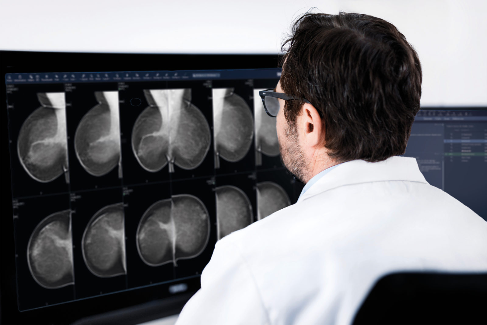
Analyzing today’s breast imaging centers, we see the need to introduce new technology and consolidate existing IT systems. However, looking back at the digitization journey of some of the first mammography departments, we see that the current demands on the evolution of IT systems are nothing new. In fact, one of the most obvious lessons learned is that for breast imaging centers to remain efficient and capable of delivering the highest quality of care, a scalable breast imaging PACS is a must. In this article, Anders Granlund, Product Manager of Breast Imaging IT at Sectra, looks back at Sectra’s nearly 20 years of experience in breast imaging workflows and summarizes the common steps our customers have taken in the evolution of their PACS. In conclusion, he tells us a few of the lessons learned along the way.
Step 1: Analogue to digital
In the early 2000s, the first mammography departments went digital with a breast imaging PACS. A pioneer in this development was Helsingborg Hospital in Sweden, one of the first mammography departments in the world to go digital. [Read their story]
Helsingborg Hospital is a part of the national screening program in Sweden and as such, had very high demands on efficiency and uptimes. The high throughput of visits—one exam every five minutes in each exam room—had to be sustained. They also required 100% uptime; this new IT system simply had to work, or else there would soon be a build-up of patients in the waiting room—many of whom had taken time off work to be screened.
Helsingborg formed one of Sweden’s first true breast unit with radiology and breast imaging integrated around the patients, however, many of the other early digital adopters did so with disparate PACS for radiology and breast imaging.
Step 2: Consolidating or integrating with radiology PACS
The first customers to digitize are now on their fourth or fifth generation of (computer) hardware and over the past decade, many healthcare providers have moved closer to the radiology department either by consolidating their Breast Imaging PACS with the radiology PACS or by integrating their best of breed breast imaging solution with the radiology PACS and a shared VNA. As pioneers in organ-oriented workflows, breast centers saw the advantage early on of having screening and diagnostic exams (often performed at the radiology department) available in the same system, to accomplish efficient multi-disciplinary team (MDT) meetings (a.k.a. tumor boards).
Step 3: Bringing in data from other disciplines and tighter integration with EMRs
A second wave of consolidation seen recently is Enterprise Image Management systems bringing data from other disciplines into the imaging system. Today, digital photography from reconstructive surgery, video recordings from the operating room, dose plans, cardiac ultrasound exams from follow-up of heart function during chemotherapy, etc. are also stored in the same ecosystem and are available at the breast radiologist’s workspace. EMR systems are more tightly integrated with the image viewers for efficient result distribution and, altogether, these improvements are highly beneficial to the cancer care treatment chain.
Step 4: Integrated breast imaging and pathology workflows
A third wave of consolidation can now be noted with the introduction of digital pathology. The interaction between the breast radiologist and the pathologist is a key component in the diagnostic workflow. With digital pathology, it is exciting to see pathology images and results also being a part of the multidisciplinary conferences in an entirely new and efficient way. Not by having a microscope connected to a different monitor or projector, but integrated digitally in the same platform as radiology, pathologists are now able to prepare MDT presentations in advance with key pathology images and radiology images side-by-side, complete with annotations and cell counts. The advent of integrated diagnostics enables more informed decisions and ultimately higher quality care. [Read the article published on healthcare-in-europe.com about Salford Royal NHS Foundation Trust in the UK and their experiences of digitizing pathology]
The trend of mergers and acquisitions also increases the requirements on efficiently distributed workflows, for example, remote reading of mammograms and digital pathology slides. New technology now makes it possible for facilities with effective distributed workflows to significantly reduce turnaround times. Access to richer information leads to actionable results earlier for the malign cases, and less worry for the negative screening cases as lay letters will be distributed more quickly than before, leading to less anxiety for the patients awaiting the results of their screening mammography exam.
Lessons learned: The benefit of a scalable infrastructure
None of the steps of consolidation, integration and efficiency improvement would have been possible had the mammography departments digitized on separate modality workstations supporting proprietary formats. Only with a true multi-modality workstation combined with an open, scalable infrastructure can these benefits be realized. The consolidated multi-modality approach has also paved the way for enriching the ecosystem with important tools for analysis of the departmental workflow, such as dose monitoring solutions and business analytics tools, both providing quality improvements, workflow optimization and statistical analysis to identify bottlenecks. History teaches us that the clinical workflows will evolve, as will technology and the opportunities to cooperate efficiently around patients and their data. With a breast imaging solution that evolves, expands and scales according to your needs and IT strategies, you are best prepared to capitalize on current and future IT innovations.
