Article

Using MRI in breast screening for women with extremely dense breasts: a first-of-its-kind study proving the benefits

Interview with one of the researchers: Wouter Veldhuis, PhD, MD

MRI in breast screening—a much-discussed topic

Utilizing MRI and breast tomosynthesis in addition to mammography, particularly for women with extremely dense breast tissue, is currently a much-discussed topic when it comes to personalizing breast screening programs. Considerable research and discussion are currently taking place to determine whether a supplementary MRI examination would be beneficial for decreasing mortality in women with breast cancer.

A recently published paper on the results of the DENSE trial[1] has shed new light on this matter and added further fuel to the debate. We met with one of the lead researchers, Wouter Veldhuis, PhD, MD and radiologist at the University Medical Center (UMC) Utrecht in the Netherlands, and asked him to describe the results and provide some insights on the study and its implications.

Dr. Veldhuis’ take on the result

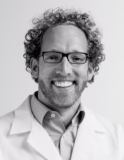
Dr. Wouter Veldhuis, University Medical Center Utrecht

Dr. Wouter Veldhuis, University Medical Center Utrecht

Can you briefly describe the results of the study and why it has received so much attention?

Our team found that, by offering an additional MRI after an initial negative mammogram (no cancer) to women with extremely dense breasts, we could detect significantly more instances of breast cancer.

A finding that is even more important was the reduced number of interval cancers[2]. Reducing interval cancers is the best-known measure for a subsequent reduction in breast cancer mortality.

Is this the first study of its kind?

There are many studies that have looked at MRI for breast screening in general. But this is the first randomized controlled study, a design that is needed to be able to also investigate interval cancers.

What do you think the biggest impact of the results will be?

The study will aid in the formulation of future breast screening guidelines. But the most important impact is that we have proved that using MRI in breast cancer screening reduces interval cancers among women with extremely dense breasts.

Why did you use interval cancers as the primary endpoint and not mortality?

Simply because it would take too long to perform such a study, at least 30 years. Interval cancers is the best known proxy measure for mortality.

When did you start the study and why were so many hospitals involved?

The preparations started back in 2009, when the team from UMC Utrecht sat down together with eight hospitals across the Netherlands. We wanted a good nationwide spread in the population. A lot of time was spent on deciding the MRI protocol (3T MRI with 23 time points), which we are very happy about today since it makes the results truly future proof.

What did you learn during the study?

All of the radiologists involved in the study met every six months to discuss the more difficult cases, and we continuously learned from each other, leading to continuous improvements.

For example, we knew that the risk of false positives using MRI is high, since it is very sensitive. But the feedback we received over the study period helped to reduce the number of false positives.

During the study, I think we doubled our precision in terms of false positives by learning to read MRIs better.

Dr. Wouter Veldhuis, University Medical Center Utrecht

Do you think any national screening programs will incorporate MRI screening for breast cancer soon?

Perhaps, but costs are also important of course. The countries most likely to adopt MRI screening are the Netherlands, the US, Sweden and Norway, but there are other factors besides costs that will come into play. We have provided data on the efficacy, and now it is up to society to decide whether it is practically possible to incorporate MRI into the programs.

What are the challenges involved in using MRI in screening?

A key issue is to make MRI screening cheap enough, and we need to reduce the number of false positives. The issue of access to equipment and radiologists must also be solved. As with the conventional screening programs, training and certification need to be set up.

Another key question to answer with respect to the cost issue is whether it would be possible to use an abbreviated protocol to handle more women per hour. In traditional mammography, we handle ten women per hour and with the full MRI protocol we can only do two, which doesn’t scale.

What other studies do you believe are needed to solve these challenges?

A cost-effectiveness analysis is currently being performed. And we are looking into whether AI might be a helpful technique for filtering out false positives to help make MRI screening affordable.
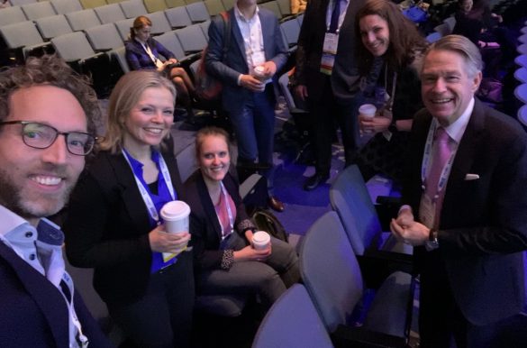
Dr. Wouter Veldhuis and team behind the DENSE study when presenting the key findings at RSNA 2019 in Chicago, USA.

From another angle

The study has received considerable attention around the world. To gain another perspective, we looked at the views of another world-leading researcher in breast cancer, Sophia Zackrisson, MD, PhD, at Skåne University Hospital in Lund, Sweden.

In an article[4], she describes her enthusiasm about the results, but she is also concerned about the number of false positive findings. It is important to wait for the final results of the second round of the DENSE trial because the number of false positives is expected to be significantly lower.

Perhaps the high number of false positives is the price you need to pay for finding these previously difficult to detect tumors. [5]

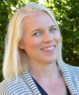
Sophia Zackrisson, MD, PhD, at Skåne University Hospital in Lund, Sweden
Dr. Sophia Zackrisson, Skåne University Hospital in Lund

Sophia Zackrisson, MD, PhD, at Skåne University Hospital in Lund, Sweden. Photo: Kim Lindkvist.

Dr. Zackrisson also highlights the need for future research to determine whether further follow-up examinations with MRI will be beneficial or not from a mortality perspective. This can be done by investigating how many of the tumors that were found in the first round of MRI were in fact slow growing and less aggressive. She states that in the DENSE study, it was unclear how many of the cancers found were life threatening and how many would be categorized as over-diagnosed, causing more harm than benefit by being found. Overall, Dr. Zackrisson is very pleased with the work carried out by Dr. Veldhuis and his team and is thrilled to continue working on improving breast cancer screening at the national level.

What it all boils down to

Altogether, the findings in the DENSE study have proven that using a supplementary MRI examination for women with extremely dense breast tissue in breast cancer screening programs will reduce the number of interval cancers, which most likely would reduce mortality. Further studies are now needed to evaluate how MRI could be included in screening programs, whether cost would have an impact, whether the protocol can be shortened and whether AI-based image analysis could help to make the incorporation of MRI practically feasible and affordable.

We are certain to see additional future research into ways personalizing screening programs in order to detect breast cancer even earlier, especially in women with dense breasts or other risk factors. Reading breast MRI exams in a screening setting will likely become more common among breast radiologists, which highlights the need for easy-to-access tools to cope with the growing volumes.


Figure 1. Table of the main results from the DENSE study[6]

References

  1.  Marije F. Bakker, Ph.D., Stéphanie V. de Lange, M.D., Ruud M. Pijnappel, M.D., Ph.D., Ritse M. Mann, M.D., Ph.D., Petra H.M. Peeters, M.D., Ph.D., Evelyn M. Monninkhof, Ph.D., Marleen J. Emaus, Ph.D., Claudette E. Loo, M.D., Ph.D., Robertus H.C. Bisschops, M.D., Ph.D., Marc B.I. Lobbes, M.D., Ph.D., Matthijn D.F. de Jong, M.D., Katya M. Duvivier, M.D., et al., Supplemental MRI Screening for Women with Extremely Dense Breast Tissue. New England Journal of Medicine 2019; 381: 2 091–2 102. DOI: 10.1056/NEJMoa1903986
  2. Interval cancers are the cancers detected in the two years after a mammogram screening, before the next mammogram.
  3. Coordinated from the UMC Utrecht in collaboration with the AVL, VUMC Radboud UMC, Maastricht UMC, Albert Schweizer, ZGT and Jeroen Bosch hospitals.
  4. https://www.dagensmedicin.se/artiklar/2019/11/28/mr-screening-minimerade-intervallcancrar-vid-mycket-tata-brost/
  5. https://www.dagensmedicin.se/artiklar/2019/11/28/mr-screening-minimerade-intervallcancrar-vid-mycket-tata-brost/
  6. Marije F. Bakker, Ph.D. et al., Supplemental MRI Screening for Women with Extremely Dense Breast Tissue. New England Journal of Medicine 2019; 381: 2 091–2 102. DOI: 10.1056/NEJMoa1903986


Author: Simon Häger, Market Strategist at Sectra

Related products