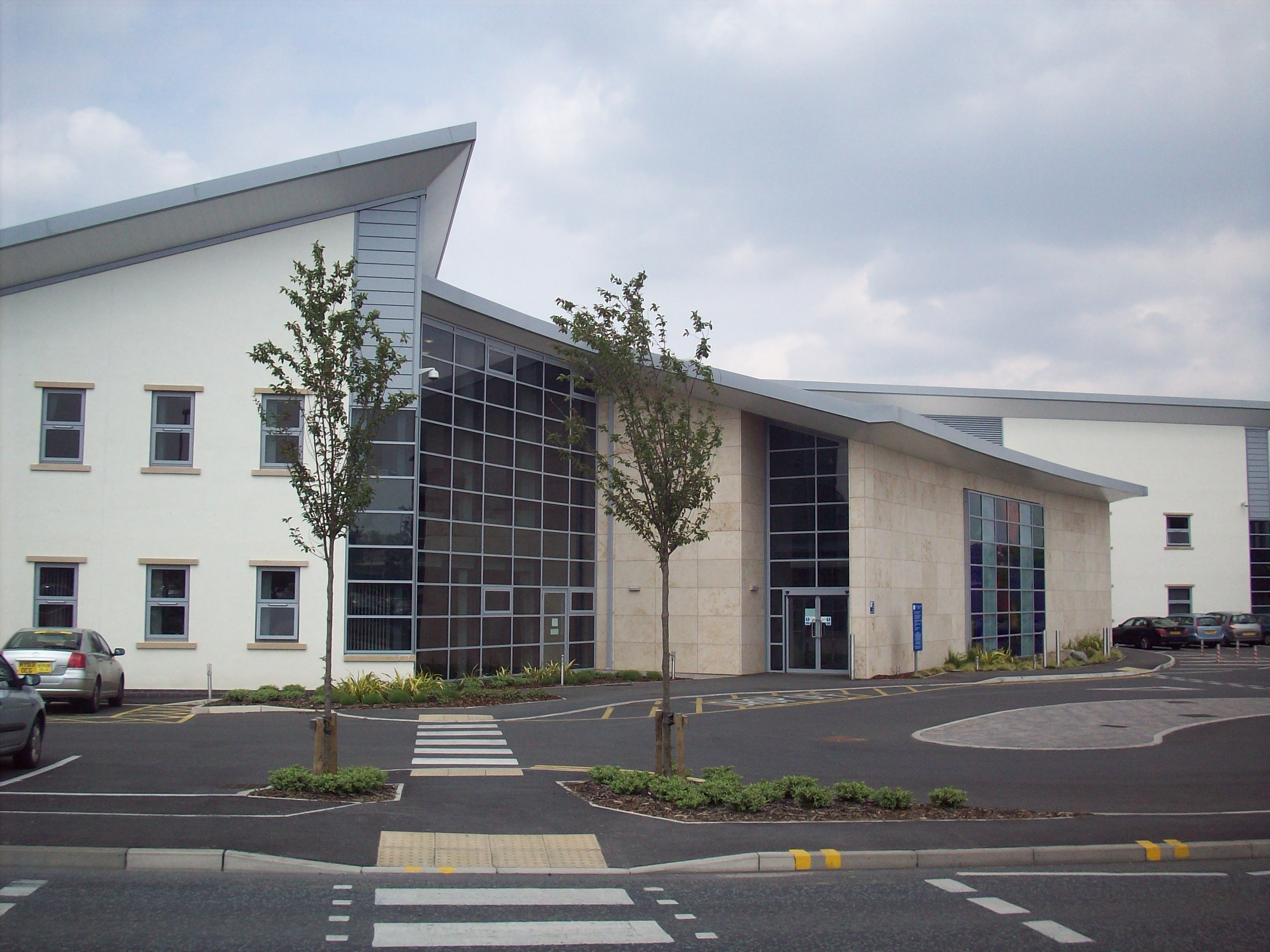
The Nightingale Centre in Manchester, United Kingdom, is Europe’s first purpose-built center for breast cancer prevention. Services provided by the center include symptomatic and screening mammography, ultrasound and biopsy. The Nightingale Centre also conducts national training programs for radiologists and radiographers.
We needed a reliable solution that could cope with the growth of the Centre, integrating with different imaging modalities, and one that offered us the flexibility to configure different user access levels. Sectra Breast Imaging PACS also offered us the web distribution solution, which means that all authorized users can view images from wherever they are.
The primary aim of the Nightingale Centre is to deliver the highest quality of care possible. As part of providing quality of care, it is also important to provide an efficient workflow for radiologists and equip them with the most advanced imaging systems and information technology available.
The Nightingale Centre is not only known for its research and training facilities, but also for its complete attention to client and patient needs. Much thought has been given to every detail within the building, from the sensitively planned layout to the calming influences of the esthetics.
Finding a PACS to complete workflows
Over the past three years, the Nightingale Centre has progressed through a number of steps to become the top modern breast screening facility it is today. Recently, the Nightingale Centre moved into new premises at Wythenshawe Hospital funded by the Genesis charity and the Strategic Health Authority.
Key challenges on the way have been integration among different IT systems and imaging modalities, support of the center’s varying workflows as well as finding solutions that are simply easy to use.
In 2006, the Nightingale Centre required a PACS solution that could integrate its existing full-field digital mammography modality (FFDM) with the National Breast Screening System (NBSS) and its local radiology information system (RIS) to acquire demographics and appointment information as well as to generate DICOM work lists. In other words, the center needed a PACS that could connect all the components together and provide the missing links of a complete workflow.
Expandability and integration capabilities
Sectra Breast Imaging PACS is designed to increase clinical productivity and provide enhanced functionality through imperceptible integration with all types of imaging modalities, information systems and infrastructures.
This was of great importance to the Nightingale Centre, as it needed a PACS with the ability to transfer images from its mammography workstation to a physician or hospital using a different vendor’s PACS or diagnostic workstation.
The expandability of Sectra PACS without performance compromise was another key factor in the Nightingale Centre’s decision.
“We needed a reliable solution that could cope with the growth of the Centre, integrating with different imaging modalities, and one that offered us the flexibility to configure different user access levels. Sectra Breast Imaging PACS also offered us the web distribution solution, which means that all authorized users can view images from wherever they are,” says Barbara Eckersley, Superintendent Radiographer at the Nightingale Centre.
Going fully digital
The first PACS installation was so successful that the Centre decided to become fully digital and bought a further five digital imaging modalities. In order to facilitate the move to an entirely digital service, the Centre invested in a major upgrade of the existing Sectra PACS to provide a comprehensive enterprise breast imaging solution. At the same time as its equipment was being upgraded, the Centre was also relocating to new premises at the Wythenshawe Hospital.
For Barbara Eckersley, it was vital that the PACS upgrade could minimize disruption, facilitate rapid adaptation by staff and clinicians, and not shut down clinical operations. “With everything else being new, the PACS just had to be problem-free and easy to use with an intuitive user interface,” says Barbara Eckersley.
In this new environment, the PACS would have to integrate with five mammography modalities from three different vendors, a number of other imaging modalities and an existing RIS and central PACS, as well as off-site screening. This required DICOM integration to the digital mammography units and the other imaging modalities, as well as continued integration with the NBSS Client Administration System and HL7 integration with the local hospital RIS.
“Sectra has proven to be dedicated to understanding and meeting our specific operational needs. Their experience of implementing integrated solutions has enabled us to reach an efficient workflow, facilitating breast screening and diagnostic service on an enterprise level,” says Barbara.
Full support for any workflow set-up
The Nightingale Centre performs both screening and symptomatic mammography, meaning that the PACS must be able to handle different workflow set-ups. Sectra Breast Imaging PACS supports independent reporting protocols, and as such, the workstation accepts single reporting for symptomatic images, yet demands double reporting with arbitration support for screening mammograms. Sectra Breast Imaging PACS also offers a solution in which different sets of privileges for users can be programmed, and it can actively use the status of an individual to determine what workflow he or she can perform.
“This is particularly important within the UK, where some of our radiographers are trained to a higher level of competency. These ‘advanced practitioners’ are qualified to report screening mammograms in conjunction with breast radiologists,” says Barbara.
The Sectra system is configured to recognize the reporting level of the individual and limits reporting access accordingly. This way, radiologists can report any images, but if an examination has been “first-read” by an advanced practitioner, the system will bar other advanced practitioners from making the “second read” and only allow a radiologist to make the “second read” or arbitrate a difference of results.
The new technology within a digital environment requires further attention to detail in the radiology work environment. Sectra recommended dedicated workstation furniture and controlled lighting to provide optimum diagnostic performance and viewing conditions. The breast service has two dedicated workstations in the reading area which have specific lighting to further assist reporting. The unit also has another three diagnostic workstations in a “hot reporting” area for use during symptomatic and assessment clinics.
Improved efficiency and diagnostic confidence
The completely integrated Sectra solution has significantly improved the workflow for the Manchester breast screening service, particularly since the NBSS does not currently supply DICOM work lists to the imaging modalities. This is instead compensated for by the Sectra Breast Imaging PACS.
“Imaging is central to the service, and the changes within the department have been positive, with better availability of images providing a much quicker, more efficient workflow,” says Barbara.
The complete system is now integrated with both NBSS and the Centre’s local hospital RIS, as well as the local general radiology PACS which is part of the “Connecting for Health” (CfH) system (CfH is the National Programme for IT which was set up to procure, develop and implement modern, integrated IT infrastructure and systems for all NHS organizations in England). The Nightingale Centre retains responsibility for all breast images it acquires, and these are stored locally within the breast unit. Back-up of the system data is provided by the hospital trust IT group. All digital imaging equipment used by the screening service is linked into the Sectra Breast Imaging PACS, so that radiologists can sit in front of a single workstation to view images from a wide range of digital equipment from numerous manufacturers.
Breast images are available to any authorized individual in the trust by means of Sectra’s web distribution technology. This allows clinicians in other parts of the hospital to view images from anywhere within the Trust’s network, whether at their own computers, surgery, oncology or projected onto large screens for multidisciplinary meetings. Web distribution is also used within the Nightingale Centre, as it provides a cost-effective way of providing images throughout the building. Standard computers within clinical rooms such as Ultrasound allow radiologists and ultra-sonographers to view a patient’s existing images before and during the ultrasound examination. Instant access to the correct images leads to improved diagnostic confidence and greatly improved efficiency.
Featured products & services
Related cases


