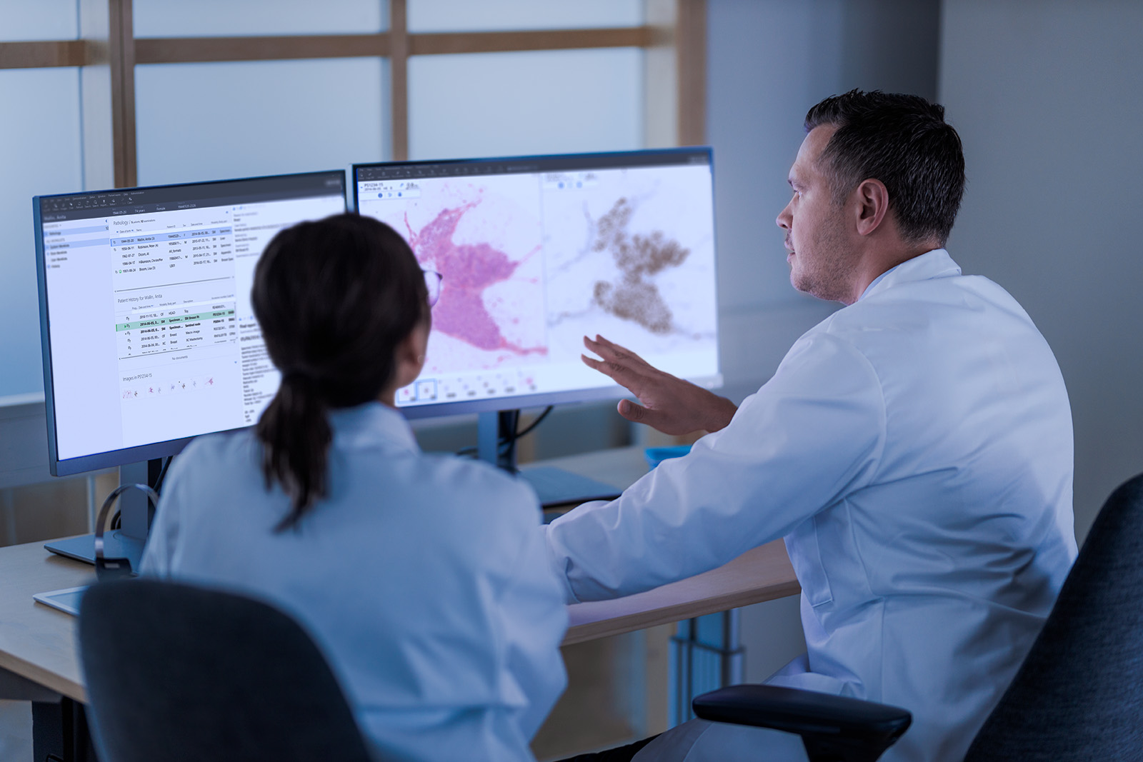
Medical imaging in the field of oncology is advancing in leaps and bounds. It is becoming increasingly easy to manage images and data, making it possible to determine with greater accuracy which treatments will improve the patient’s quality of life and how those treatments can be applied in the most targeted way possible. It is also becoming clear that, in some cases, it’s preferable not to provide any treatment at all.
Sectra discussed these developments with two leading hospitals that both collaborate on a multidisciplinary basis to further improve their patients’ treatment. We spoke at length with a number of thought leaders from AZ Delta Hospital and University Medical Center (UMC) Utrecht about their views on the relationship between medical imaging, oncology and IT, and the improvement of patient care.
We are aware that pathology and radiology are very similar in terms of processes and content. We both advocate for a more integrated form of diagnostics and have been looking for opportunities for improved integration and collaboration at a technical and content level for some time.
Pathology and radiology collaboration at UMC Utrecht
Prof. Dr van Diest is Head of the Department of Pathology at UMC Utrecht and is focused on digital pathology and artificial intelligence (AI). Dr Veldhuis is an oncological radiologist and co-founder of UMC Utrecht Radiology-AI start-up QuantibU. Prof. van Diest explains: “We are aware that pathology and radiology are very similar both in terms of processes and content. We are both advocates for a more integrated form of diagnostics and have been seeking better opportunities for a long time in terms of integration and cooperation at a technical and content level.”
When it comes to the digitisation of medical imaging and workflow optimisation, pathology and radiology alternate, as Dr Veldhuis explains: “We [radiologists] already had digitised procedures, but if you look at structured reporting, we are lagging far behind. Attempts have been made to structure reporting, especially for prostate and breast imaging. A system like PALGA, which pathologists use, would also be very useful for radiology. Prof. van Diest adds: “Improving the workflow requires a local champion. Someone has to take this on. You have to start small and then grow.”
Trends in digital pathology
Digital pathology mainly involves digitising tissue sections and setting up digital workflows. Prof. van Diest continues: “Sectra provides us with access to the PIE (Pathology Image Exchange) platform supporting the digital exchange of images between laboratories for consultations, peer supervision and trials, and possibly also for research and training purposes. As digitisation increases, there is a greater need to collect diagnostics on a reciprocal basis. In addition, regional networks are emerging where the PACS is shared.”
We have AI algorithms running that know when to start, so that a result is available when I start reading. This is very important for patients and we will make great strides in this field in the future.
In terms of AI implementation, the workflow will change in the period ahead. “Ideally, you automate the workflows in such a way that processing operations are already finished when we receive the image, for example, if the system takes us directly to the specific tissue section where an abnormality can be seen. This will save time.”
Trends in radiology
A great deal of progress has been made gradually in the field of radiology: “If you look back at the last 15 years, enormous technical progress has been made in MRI and CT techniques. Patients are also scanned much more often before or during treatment, or as part of investigations. As a result, we are increasingly receiving more detailed images. We are slowly starting to look at this more in quantitative terms. Specialists also speak to each other more often, which increases mutual understanding and know-how,” says Dr Veldhuis.
He continues: “We have AI algorithms running that know when to start, so that a result is available when I start reading. This is very important for patients and we will make great strides in this field in the future. One example of an area which is now attracting a lot of attention is prostate cancer where we are using AI to tackle it in a structured way. Our software reads along with us, segments the prostate, calculates the PSA density and indicates where lesions are likely to be. If you approve it, you get the volume of the lesion. In this way, you are guided through the entire process. We now have a dedicated viewer that specifically helps us with the prostate. Ideally, you want to have that kind of interaction in the PACS as much as possible so that it always works and is stored in the same way.”
Aspects for improvement in the short term
According to Dr Veldhuis, progress is being slowed down by the fact that we do not really have a system for structured reporting yet. “For example, there is no support for the multi-faceted nature of reporting. Suppose someone has viewed something and then an expert opinion comes out about it, or extra clinical information becomes available that changes the interpretation of the images, then I want to be able to record this in an effective way. We can learn am awful lot from a system that supports more readers, measurements and measurement variations. More knowledge provides more nuances and you want to be able to save this using [intermediate] steps. This shows where we often go wrong, what we can learn from and what we are best at.”
It will take another 10 years before we really feel an impact, because more cooperation is needed, along with more data that you can store in a structured way.
The external and internal exchange of images is another area where there is potentially a lot of room for improvement. “Images are often received from other hospitals, including radiology reports, only in the form of a scanned image, which I cannot search and have analysed by a computer. And if I produce an addendum, it is not automatically transmitted back to the original hospital in a structured way,” adds Dr Veldhuis. “However, the patient often goes back there for part of the treatment and follow-up. The data exchange process can also be improved internally. For example, there is still no structured way of reporting to help improve the interpretation of images based on a discussion in an MDC (multidisciplinary consultation), except for an [input] field in the EHR. This makes it much more difficult to learn from it and know for sure that important details are being followed up properly.”
Looking ahead: further digitalization and cross-disciplinary cooperation
Within five years, Prof. van Diest and Dr Veldhuis hope they will have improved the workflow even more, but, according to Dr Veldhuis, “it will take another 10 years before we really feel an impact, because more cooperation is needed, along with more data that you can store in a structured way. What is currently available in terms of AI still has only a limited impact. It’s useful, but is not the same as making a diagnosis.”
Prof. van Diest mentions a number of major advantages favouring the future of imaging:
- First of all, complete digitization of all pathology laboratories in the Netherlands is required because you need to have the infrastructure first. At the moment, about half of the pathology labs have a digital infrastructure.
- Next, you need to ensure permanent storage of images. We are one of the few pathology departments that keeps them indefinitely. This poses a challenge in terms of costs, technical aspects and infrastructure if the images disappear at some point while you have done your best to digitalise everything. These images also still need to be integrated into various patient information systems.
- Furthermore, regional networks must be set up so that shared or even joint systems for digital diagnostics can be created. The PIE platform could be expanded for diagnostic purposes.
- Also, an ever-increasing number of AI algorithms will be implemented locally or centrally.
- For training and research purposes, a central image archive can be created.
- Finally, we can work more on a cross-disciplinary basis, while integrating disciplines in the process.”
Using images to provide targeted treatment at AZ Delta Hospital
Our collaboration covers the entire process, from the initial complaint or increased PSA value during a doctor’s appointment to potential treatment, collection of PROMs and PREMs studies, and all the steps in between involving imaging and reporting. This enables us to optimize the subprocesses involved to achieve a better result.
Intensive cooperation during the entire diagnostic and treatment pathway
Dr De Smet is head of radiology with a subspeciality in cardiac and urological radiology at the AZ Delta Hospital in Roeselare and a particular interest in prostate cancer. Together with urologist Dr Goeman, he performs transperineal NMR-guided prostate biopsies, together with other procedures. Dr De Smet explains: “Our cooperation covers the entire process, from the initial complaint or increased PSA value during the GP visit to potential treatment, collection of PROMs and PREMs studies, and all the intermediate steps involving imaging and reporting. This enables us to optimize the sub-processes involved to achieve a better result.” This includes both the actual, hard outcomes and the subjective assessment with regard to the patient’s quality of life.
Drs Goeman and De Smet work closely with Prof. Dr De Jaeger on the data processing side of things. As Prof. De Jaeger explains: “As an engineer, I try to make what the doctors require fit into an elegant process. This then makes it clear where there is an interchange and where improvements can be made.” He believes that the hospital of the future is a place where specialists will not only collect patient data, but also merge it to thus learn from it. “In the past, you had a photo to work with, now you have a dynamic report. You know what you should or shouldn’t do; it’s treatment-defining.”
Role of MRI in prostate cancer
Dr De Smet continues: “Once a referral has been made, an MRI is an extension of the first-line examination. The images are required to assign any prostate injuries a PI-RADS classification.” In his view, the MRI fulfils two roles: firstly, it is a pre-biopsy gatekeeper as part of the risk analysis and, secondly, it is a navigator when a biopsy is still needed.
“This kind of MRI, combined with other parameters such as the PSA value and a physical examination, enables us to determine whether a biopsy is desirable. We not only want to learn from a large cohort [as with prostaatwijzer.nl], but also from our own patient population.” An MRI ensures that we can do this as specifically as possible. “A focal biopsy is different from the usual standard biopsy.” Conversely, in some cases, a standard biopsy is carried out based on the data obtained, even with an MRI not showing any obvious injury.
We don’t want to cause any harm by treating patients for a condition that isn’t life-threatening and won’t affect their quality of life. In order to make even better decisions, we carry out data analyses.
Treatment is not always required
During multidisciplinary cooperation, structured and standardized reporting is of paramount importance. As Dr De Smet explains: “This allows us to become smarter when using this data, to ensure that we learn from it and perform better in the long term.” All the information collected can be used to train algorithms and obtain the most personalized result for the patient. “Precision medicine then enables us to better offer the care which patients expect.”
The team is going through a transition where reporting is done in a more integrated way and patients are being treated unnecessarily even less often. As Dr Goeman sees it: “Not treating patients unnecessarily is the most important outcome of this cooperation. We feel it is important for us to be able to offer patients the best possible quality of life. If someone is not treated, it does not mean that they have been cured, but we can also say that they should just not be treated right now. We don’t want to cause any harm by treating patients for a condition that isn’t life-threatening and couldn’t affect their quality of life either. To ensure that we are in an even better position to make these decisions, we carry out data analyses.”
Dr De Smet adds: “The development phase we are now going through is all about high-precision diagnostics and actually knowing what is going on, so that any [potential] treatment is provided as locally as possible.”
Beyond the PI-RADS score
The first benefit gained from optimizing the workflow is an increase in performance and continuity. Prof. De Jaeger explains: “The size of the data files is not too bad at the moment because the data is in text form. It comprises numbers and values. The image is now being converted into a PI-RADS score, but it would be [even] better to be able to create a kind of convolutional network, in which characteristic properties are retrieved directly from the image and combined with the PSA data from the laboratory. This then gives you an even more objective system. This requires more storage and data processing, but this is part of version 2.0. We will initially continue with version 1.0, using the relatively simple data. In Flanders, it is also possible to use Flanders’ subsidized supercomputer centre for performing computationally intensive tasks.”
A system that continuously learns
Prof. De Jaeger continues: “Ultimately, the outcome is the most important thing for the patient. If we collect the data in a structured manner, we can train a separate model and predict the outcome of using treatment A or B. If this is not clear, there are also discussions where we can provide doctors with much more data-driven support. The intention is to make judgements a lot more objective.”
Dr Goeman adds: “We want to extract as much data as possible that is still ‘hidden’, or analyse it in such a way that it guides us in a particular direction.” Prof. De Jaeger adds: “If we can retrain the system on a weekly basis, it will first require data preparation, but then you will have a really powerful system that continuously learns later on.”
AI reorganises the radiologist’s tasks
Images will probably be interpreted with the naked eye less in the future, at most during the test phase for an algorithm. Dr De Smet explains: “This is proceeding hand in hand with a transition that is taking place in radiology, with a shift from visual interpretation to a new set of tasks, which involves a certain amount of health technology assessment, in which you select and retrieve algorithms based on performance [for the use case]. The next step is to monitor the quality of the generated data. Based on much more in-depth analyses, you provide consultancy to the referrers and about the process which the patient will go through.” Health technology assessment, quality management and clinical consultancy are new core tasks for radiologists, in which AI frees up the necessary time, thus allowing radiologists to study the content further. This renewed focus will only help the role of the radiologist become more relevant.”
Targeted treatment and multidisciplinary cooperation
AZ Delta is not lacking in ambition when it comes to prostate cancer. Dr De Smet explains: “Given the current rate, a great deal will have changed in five years’ time. I hope to perform our first focal treatment within a year.” Prof. De Jaeger initially foresees an acceleration of PROMS studies as a basis for training algorithms.
In addition to ensuring that data processing is as well structured as possible, the interviewees recommend good cooperation between data scientists and medical specialists. In the long term, this requires a necessary consolidation in the sector. Dr De Smet continues: “I think this is how the future will be for all fields. If you take the example of prostate cancer, incidence is only increasing – the only form of prevention is not to grow old. It is a major problem for men, which is why we have to adopt this new way of working.”
Multidisciplinary cooperation requires open communication and coordination of the various areas of expertise. As Dr Goeman explains: “It is a sign of the times and part of the educational spirit that urologists and oncologists, for example, look at the same case differently. Thanks to imaging, we now have a better picture of how a tumour behaves. Urologists sometimes still look too often at biomarkers such as the PSA value, which are kind of snapshots. We have now seen that we can work together more with radiologists and what you can do with this data.”
Prof. De Jaeger concludes: “I think we have a pioneering role to play. The more data there is, the more accurate the result [can be]. It’s good if more people are involved, as we are ultimately doing this for the patients.”
Featured products & services
Related cases


