Orthopaedics

Plan before surgery and cut time spent in the operating room

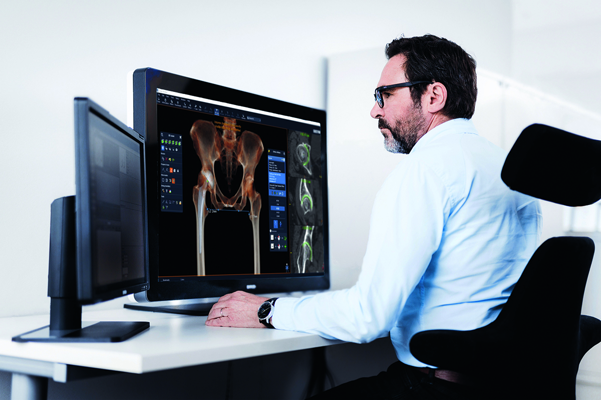
Modern planning systems with 2D and 3D images lets orthopaedic surgeons and MSK radiologists diagnose and plan orthopaedic procedures—for instance knee, hip and complex trauma surgery—with greater accuracy and confidence. With increased precision in planning and more advance preparations, surgeons and operating teams can reduce stress, save time, and minimize risk during surgery. For the patients, planning reduces the risk of postoperative complications. For hospitals, it creates a well-documented workflow to meet regulatory demands and improves the teaching facilities.

Selection of common use cases

Visualize trauma structure and improve operating efficiency

Pre-op planning on 3D images improves planning of such complex cases as multiple fragment traumas and deformities. By visualizing trauma structure, you can diagnose a fracture without first opening the patient. This minimizes the risk of surprises and stress during surgery, which also shortens the time spent in the operating room.

3D pre-op planning allows trauma teams to correctly diagnose and plan for surgery, to save valuable time during operation, and to improve the quality of diagnosis and the treatment plan. In addition, planning on 3D images facilitates communication in the surgical team, further improving operating efficiency.

Learn more

Increase accuracy in joint replacement with 3D planning

Sectra’s 3D Joint Replacement solution lets surgeons easily plan complex arthroplasties. The 3D views and dedicated orthopaedic tools allow for increased accuracy in implant sizing, angle measurements and surgical approach. Surgeons can now study the patient’s anatomy, and plan for surgery, in ways that are simply not possible using today’s standard 2D images. This can ultimately lead to less time in the operating room and improved surgical outcomes.

With Sectra’s 3D Joint Replacement solution, surgeons can gain advantages that prove highly beneficial for their patients. The intuitive tools enable a quick learning curve, while template and angle measurement functions speed up the process of planning through simultaneous use of 3D and MPR views. 3D joint planning also seamlessly removes potential calibration issues and errors, as CT data visualizes true size automatically, improving accuracy in implant selection even further.

Learn more
Improve planning before spine surgery

Sectra’s 3D spine solution allows surgeons to plan more efficiently for cases involving pedicle screws.  The solution considers each pedicles’ unique anatomy when determining placement and sizing of individual screws.  From standard fusions to complex scoliosis, Sectra is there to help you plan with confidence.

Learn more

Print your orthopaedic cases in 3D

Printing orthopaedic cases in 3D offers several benefits. A printed 3D model allows for further surgical planning after a pre-operative plan has been created and can be used for educational purposes, as a template for pre-bending implants for an upcoming surgery or even as a keepsake for the patient or surgeon.

Learn more

Easy pre-op planning with high precision

With the right orthopaedic tools for 2D pre-op planning, you can enhance the preparation of orthopaedic surgery of all body parts—including hip, knees, shoulders, spine, as well as in trauma cases.

With a few simple clicks, you can select a prosthesis with a perfect fit for the patient from our templates database. This results in high precision and efficient performance in your pre-operative planning process.

Learn more

Find out if an implant is loose with Sectra IMA

With Sectra Implant Movement Analysis (IMA), surgeons can find out if an implant is loose with the help of a CT scan. Patients who experience chronic pain after an implant surgery can thereby avoid unnecessary risks, such as infections and complications that may be caused by an avoidable surgery, if it turns out that the implant is not loose.

Learn more

Simplify and speed up the validation of new orthopaedic implants

Sectra’s CT-based implant Micromotion Analysis (CTMA) simplifies and speeds up the process of validating new orthopaedic implants in vivo. CTMA uses CT images taken up to one or two years after implantation to measure 3D motion of an implant or bone fragment on a submillimeter scale. This information provides the surgeon and implant manufacturers with a strong predictor of the long-term survival of the implant.

Learn more

Increase efficiency using innovative tools in 2D and 3D

Fast access to advanced measurements within the field of MSK radiology will save valuable time for both orthopaedic surgeons and radiologists. With Sectra’s toolbox MSK toolsfor MSK radiology, these measurements can quickly be performed using intuitive click guides displayed in 2D as well as 3D—adding an extra layer of understanding and supporting meaningful discussions. Having access to all the tools directly in the PACS will streamline the workflow, with time savings and faster diagnosis as a result.

Learn more

Reduce intra-operative complications

Digital images allow digitalized pre-operative planning, also called templating. Digital templating offers the ability to solve the scaling problem known to affect traditional templating with analogue plain images. Pre-operative planning is an essential part of the surgical procedure, and is of significant value to successful arthroplasty. It makes it possible to plan for the size of components needed, and to achieve correct positioning and orientation of the components to equalize leg length and reduce intra-operative complications.

Learn more (PDF)

2,500 reasons to trust us

Meet us next at

26
Feb
2025
to
01
Mar
2025
ECR 2025
Event | Radiology imaging | AI in medical imaging | Breast imaging | Cardiology imaging | Digital pathology | Enterprise imaging | Enterprise platform | Medical education | Ophthalmology imaging | Orthopaedics | Sectra One Cloud | Share and collaborate