If not already implemented, establishing an enterprise imaging (EI) system for managing medical content is in most healthcare providers’ plans. A common way of reaching the goal of establishing such a foundation is to expand the existing radiology picture archive and communication system (PACS). In doing so, healthcare providers can leverage established electronic medical record (EMR) integrations and PACS infrastructure. In essence, adding additional ologies to an existing platform reduces both cost and complexity since the number of vendors and systems can be minimized.
Adding cardiology as one of the first ologies — in addition to radiology — has been a proven recipe for demonstrating early wins. Aside from the financial benefits of cost sharing, this approach has led to reduced turnover times and more satisfied end-users, but only if the system meets the clinical needs of the cardiologists.
But adding cardiology is not without its challenges. The absence of standards, security issues and a lack of enthusiasm are some of the possible hurdles mentioned in a report from Signify Research[1] published in 2019. Showing early wins and addressing these challenges are necessary for a successful implementation.
This article will explain why you should add cardiology as one of the first ologies to your enterprise imaging system, and what functionality the system will need to provide to satisfy user requirements while leveraging platform synergies.
Adding cardiology as one of the first ologies
The aforementioned Signify Research[2] report states that when expanding the radiology PACS to enterprise imaging, it became clear that there was some low-hanging fruit to be had by adding cardiology as one of the first ologies. According to the report, EI project implementation can extend over a period of ten years or more, and that the “lack of early success for EI leads to early stifling of many projects”. Therefore, demonstrating early wins will catalyze further momentum for additional investments and fuel the necessary organizational and cultural change.
The main reason for cardiology being suitable to add early is that the specialty is becoming more image intense and requires support for facilitating tighter collaboration with radiology. Even though the addition of cardiology multimedia to the EI system requires a migration, it quickly pays off in the form of reduced costs and better clinical benefits.
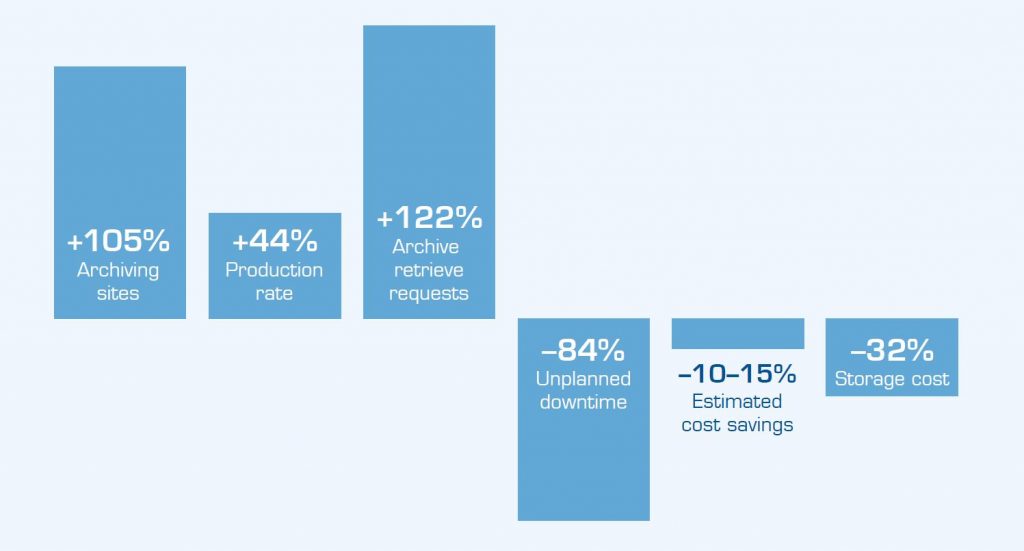
This was, for example, the solution implemented at the University Hospitals of Cleveland in 2015. A study[3] published in 2019 shows the remarkable results achieved by adding cardiology to radiology as a first step toward enterprise imaging. This reduced downtime by 84%, storage costs by 32% and overall IT infrastructure costs by 10–15%.

Post-implementation vs. pre-implementation status of the enterprise imaging system at the University Hospitals health network[3].
The increased use of cardiology imaging
To meet the requirements of cardiology end-users, it is important to have some background information on when and how imaging is used in cardiology.
Cardiovascular diseases (CVDs) are the most common cause of death globally[4]. Cardiac imaging is used in the identification of those at highest risk of CVDs, in diagnosis and in the treatment steps. Technological advancements within cardiology imaging lead to fewer invasive interventions, thereby justifying the use of more cardiology imaging.[5]
Different cardiac imaging techniques and modalities
A variety of clinical symptoms may arouse suspicion of CVDs. Various cardiac imaging techniques are often required in order to establish a diagnosis and proper treatment. From a cardiologist’s perspective, the choice of cardiac imaging modality depends on:
- The disease being investigated.
- Individual patient characteristics.
- The accessibility of tests.
For example, assessing dyspnea and investigating coronary artery disease both require cardiac imaging. And imaging is also common in the diagnosis of cardiomyopathy, and structural or congenital heart disease.[6]
The various modalities used include:
- Echocardiography
- Electrocardiography (EKG/ECG)
- Fluoroscopy in the catheterization laboratory
- Nuclear myocardial perfusion imaging
- Cardiac computed tomography (CT)
- Magnetic resonance imaging (MRI)
All of these have various advantages and disadvantages. Often, at least two of these examination types are required to obtain a comprehensive overview of a patient’s cardiac health.[7] For example, cardiac MRI is used as an adjunct to other imaging modalities when further clarification is warranted. Another example is echocardiography, which can provide the required clinical information without radiation exposure but is often used in combination with other imaging techniques to provide a broader picture.[8]
Also noteworthy is point-of-care ultrasound (POCUS)[9] — one of the fastest growing cardiac imaging techniques. POCUS has during the latest years become an important additional source of revenue and will probably keep growing rapidly in use. These images and videos are often acquired outside of the radiology department.
The list of modalities is long. And an EI system must support all of them to be able to store and enable content for diagnostic review.
Greater need for cardiology–radiology collaboration
Cardiology imaging is producing more content than ever before and this needs to be stored in a cost-efficient and consolidated manner, integrated into clinical workflows. The increase in cardiac imaging has also led to a growing need for collaboration between radiology and cardiology. The common view among cardiologists and radiologists is that this can be a challenge. The problem is exemplified by the fact that only a few successful and truly collaborative radiology–cardiology IT systems exist on the market.[10]

