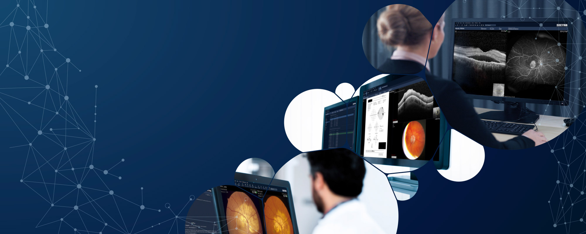
Lessons learned from radiology, cardiology and other image-rich specialties, is that physicians were wasting a lot of time working across multiple workstations. They were at a disadvantage when they couldn’t access changes in images and data, side by side, over time and confer on images and data with colleagues in an efficient way.
Why has ophthalmology been overlooked for so long when it comes to modern IT architecture? Partly because eye care is more of a “one-stop shop,” Svärd notes, where the same department or clinic is responsible for seeing the patient, performing various imaging studies, interpreting the result, and then planning and executing treatment.
But being separate is no advantage and that’s why one of the largest U.S. healthcare systems approached Sectra when they needed a new eye care imaging and data management system. The healthcare system was facing end-of-life with its ophthalmic imaging solution. They found the maturity and scalability of the Sectra Enterprise Imaging platform appealing but also recognized a need for strengthening the ophthalmology specific capabilities. Guided by their key clinical users, Sectra managed to close this gap.
Today that healthcare organization uses Sectra’s Enterprise Imaging solution to manage 1.8 million eye care studies per year.
This momentum is stirring an evolution. As a result of enterprise imaging, ophthalmic imaging is on the verge of an overhaul that will make the field more organized, standardized, and operationally and economically more efficient.
Tools for easy image review
“Our enterprise imaging solution contains more than just image storage and sharing,” Svärd says. “It also includes a diagnostic application with operational tools and functions that allow for easy comparison and analysis of images, helping ophthalmologists do their jobs better and faster. And it’s scalable for offices, departments and healthcare systems.”
Here are some of the most utilized tools and features for common ophthalmic imaging modalities available in Sectra Ophthalmology Imaging:
- Optical coherence tomography (OCT). OCT scans are presented as a scrollable stack together with a smaller fundus image with reference lines for orientation. Retinal images also allow a higher axial resolution, making it easier for physicians to see and analyze the layers within the retina.
- Fundus photos. For fundus photos that capture color images of the retina, Sectra Ophthalmology Imaging offers various color adjustment options, including the ability to show individual color channels. Measurement tools make it easy to measure findings to calculate optic cup-to-optic disc ratios, prepare for photodynamic therapy and track lesions over time. Measurements performed in standard compliant ultra-widefield photos will compensate for the large distortion in the periphery caused by the large curvature of the retina.
- Visual field test. For visual field tests that look for weakened vision or blind spots across the entire retina, the ophthalmology module offers a tool to quickly assess a patient’s progression over time by quickly and easily moving through consecutive images from previous visits.
The diagnostic application also allows ophthalmologists to easily access MR and CT images along with viewing tools, rather than having to go to a separate workstation to view them. They also can view images from niche devices such as ultrasound and corneal topographers can utilize tools to review the output. For all modalities, the eye care provider may lock on the left or right eye to be sure they’re consistently focused on just one at a time.
Side-by-side comparison with a single sign-on
When an ophthalmologist wants to review images from two different modalities, Sectra Ophthalmology Imaging allows them to view the images side-by-side on one screen, all with a single sign-on. This is one of the features ophthalmologists have appreciated most: the ability to pull up all relevant images from all modalities in less time, and with fewer clicks.
“In many practices, the first thing an ophthalmologist will do in the morning is to start logging in to all of their different imaging applications,” Svärd says. “If they want to review two different types of studies, they’ll need to flip back and forth between different applications to review them. One enterprise platform and a single diagnostic application changes all of that.”
