Faculty of Dentistry at the University of Oslo

Dental radiology is following the enterprise imaging trend

We spoke at length with lecturer and physicist Gerald Torgersen along with Linda Arvidsson and Ulf Riis, maxillofacial radiologists at the Department of Maxillofacial Radiology, part of the Institute of Clinical Dentistry at the University of Oslo’s Faculty of Dentistry. They have used Sectra’s Enterprise Imaging solution for dental radiology since 2007 and agreed to share their experiences as pioneers in this area. They also agreed to give their opinions on the pros and cons that may be valuable to know for others planning to go down the same route.

Filip Klintenstedt, global product manager for enterprise image management and VNA, also shares his views from a global perspective on the trend of including dental imaging on the enterprise imaging agenda.

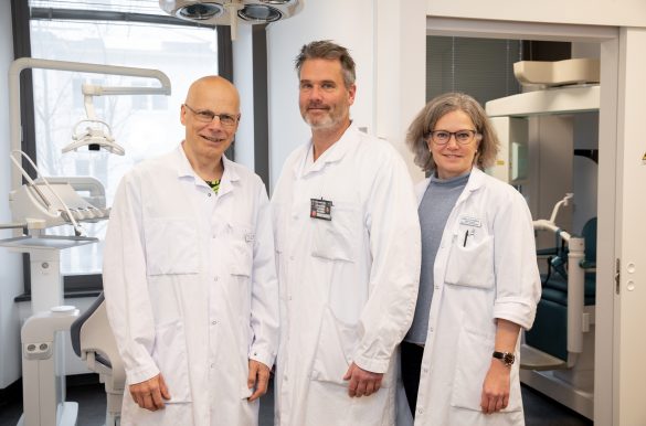
Lecturer and physicist Gerald Torgersen along with Linda Arvidsson and Ulf Riis, maxillofacial radiologists at the Department of Maxillofacial Radiology, part of the Institute of Clinical Dentistry at the University of Oslo’s Faculty of Dentistry

More and more healthcare providers are expanding their radiology PACS into an enterprise imaging (EI) system. This means going from using separate diagnostic imaging systems for each ology to a single consolidated system. The EI system is responsible for managing the storage, workflow and review of images, not only for radiology but also for pathology, cardiology, ophthalmology and, in many cases, dental imaging. This creates huge benefits from both a clinical and an IT consolidation perspective.

The shift towards EI has led to greater interest in also including dental radiology and dental images. However, dental care is normally organized separately from traditional healthcare, with dental imaging performed at outpatient dentist practices (normally dental x-ray only) and maxillofacial radiology (more advanced modalities such as CT and CBCT) performed at hospitals. This has resulted in dentists and specialized dental radiology departments using separate systems, forcing radiologists to shift between two often unsyncronized applications for each patient to get a complete overview.

Being able to import, manage, and view images from all different modalities is key to unlocking the full potential of a digital system.

Gerald Torgersen, lecturer and physicist at the Department of Maxillofacial Radiology, part of the Institute of Clinical Dentistry at the Faculty of Dentistry, University of Oslo

Utilizing the EI system for managing dental imaging provides a cost-efficient opportunity to bridge this gap and improve the tools for both radiologists and dentists, while also improving IT infrastructure, system robustness and security.

Oslo gets to the root of the problem with a common system

Gerald, who also works with IT at the Department of Maxillofacial Radiology, explains that when the institute began planning to replace dental film around 2005, they needed an archive that could include all types of images. Back then, and still today, the most common setup for maxillofacial radiologists was to use a PACS or an EI system for advanced radiology such as CT, MRI and CBCT studies, together with the separate dedicated dental system used at the outpatient clinics where plain x-ray dental and panoramic images were taken. Gerald explains that they saw this setup, which involves using two unsyncronized desktops, as risky from a patient safety perspective. He adds: “Aside from being a patient risk, it is also very inconvenient for the radiologists having to switch context to get the entire patient overview.”

The radiologists at the department were positive from the start. “Clinically, it makes sense to view all images in the same system since dental and maxillofacial radiology are similar to all other types of radiology. We review these images in a similar way and have almost the same needs for diagnostic tools,” Linda says. “The only difference is that as maxillofacial radiologists we focus on the teeth and jaw, which sometimes requires special third-party applications for analysis, segmentation or treatment planning.”

Gerald highlights another important reason why they went with Sectra’s Enterprise Imaging Solution, namely that dedicated dental systems seldom support the full DICOM standard. “Being able to import, manage and view images from all different modalities is key to unlocking the full potenial of a digital system.”

Based on these two drivers, they decided to manage both dental radiology and more advanced imaging in Sectra’s Enterprise Imaging Solution. According to Ulf, having all images, including clinical photos for treatment planning, in the same system is still one of the greatest benefits.

How it is used today

In contrast to the students, the radiologists and radiology residents work directly in Sectra’s Enterprise Imaging solution. Images are accessible immediately after they have been quality assured by the radiographers or instructors. “Having a single archive and system for all images makes it easy to work together with other radiologists, dentists and students—regardless of which department you’re in,” says Ulf.

When it comes to usage, Gerald highlights the fact that the high imaging volumes they produce, together with the hundreds of simultaneous users, place high demands on any IT system. He says: “The stability of Sectra’s solution has been key for us. Having hundreds of users logged in at the same time does not affect the performance at all. This is another benefit of using a system designed for very high demands and large institutions such as Sectra.”

Pros: Access to all images in one system

As radiologists, both Ulf and Linda agree that having access to all images in the same system is the biggest benefit of including dental radiology in one EI system. “Today, many other dental schools use separate systems for dental images and advanced radiology images,” Linda says. She adds: “Such a setup may cause inefficient workflows and frustration among users—things that we have avoided.”

Ulf explains that without the Sectra Enterprise Imaging solution, radiologists and dentists not only have to shift between two different systems, but also use different viewers for each brand of modality. “Having all images in a single hanging significantly improves my work compared with having separate systems and multiple viewers,” he says. “A single system also leads to higher patient safety,” Ulf continues. “Having access to all images from every modality, including both current and prior exams, is a kind of quality assurance.”

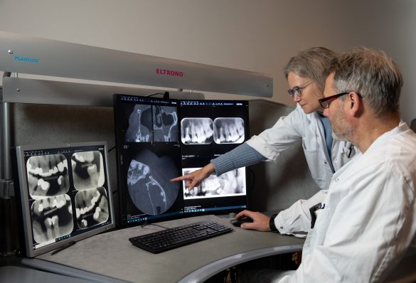
Ulf and Linda using the Sectra Enterprise Imaging solution for dental radiology. 

As an academic institution, the Faculty of Dentistry receives a lot of referrals from other private and public dental clinics across the country—both for reporting on complex cases and for providing second opinions. “Managing such a distributed workflow and exchange of images nationwide is something that Sectra’s Enterprise Imaging solution can handle very well since it is built for similar workflows in radiology,” says Linda. “Using one EI system also makes it easier when we request priors from other dental clinics and radiology departments, since it uses a standardized DICOM-based method for sharing images,” she says.

Gerald also highlights some of the benefits from an IT angle: “The system’s stability and security are a major advantage. Sectra’s solution is very robust and designed with IT security in mind.”

The best-of-suite strategy that comes with an EI system means that the number of IT solutions can be drastically reduced, which cuts costs and improves IT security.

Filip Klintenstedt, global product manager for enterprise image management and VNA at Sectra

Cons: Best of breed vs. best of suite

When asked about the biggest disadvantage of dental radiology using an EI system, Linda brings up the issue of user habits. Dentists who are used to working in a dedicated dental system might find it hard to switch to a system that functions differently. “Not necessarily in a worse way, but different to what they are used to,” she says.

Ulf adds: “Of course an EI system will never be as tailored for dental imaging as a dedicated dental system, but we have solved that by creating tight integrations with third-party systems.”

Gerald adds: “It is hard to come up with disadvantages of using Sectra, but ironically the biggest disadvantage is that we receive so few complaints in comparison to our EMR that we haven’t found enough reasons to develop it to its full potential. If our users weren’t happy with the system, we would have done more to extend its capabilities, but there has been a lack of urgency in this regard.”

Drilling deeper into the most commonly used functionality

Filip explains that Sectra’s key strategy for its enterprise imaging solutions is to make the “bread and butter” functionality that is used most frequently as efficient and intuitive as possible. “This saves the most time in contrast to sub-optimizing specific advanced features,” he says.

One example of this is the MPR functionality, which Linda explains is one of the most used and appreciated feature among their maxillofacial radiologists. She says, “It allows them to easily view different and customized planes in the images.” Linda continues: “Standard sizing tools such as measuring distances and angles are also used regularly. What is important is that these are quick to access and easy to use.”

Ulf mentions that the 3D reconstruction function is popular when demonstrating images to those who are not used to viewing image slices, particularly since it makes it easier to see complex cases. He says: “It serves a nice pedagogical purpose when showing a case to other specialists, students and the patients themselves.”

For functionalities that are highly dentist-specific, such as advanced tooth implant or orthodontic treatment planning, Linda and Ulf say they use third-party applications integrated with Sectra’s Enterprise Imaging solution. Gerald explains that these integrations are made via standardized APIs and are easily accessible by right clicking on the images where they need to be applied.

Being able to seamlessly integrate advanced applications is something that Filip describes as important for the entire enterprise imaging strategy. It makes it possible to meet the needs of various specialties by establishing tight integrations with advanced tools within one user interface. “The user should never have to switch context and lose focus,” he says.

When it comes to teaching, Gerald says that the teaching file functionality is the most commonly used and appreciated. It allows users to add specific tags to cases in order to create repositories for educational or research purposes. This functionality also allows users to easily export images to presentations for educational use.

Overall, the tools most frequently used by maxillofacial radiologists are very similar to those regularly used by other specialties within radiology. Sectra’s Enterprise Imaging solution therefore fits the needs of maxillofacial radiology well. When it comes to dental-specific advanced tools that are not available, Ulf and Linda explain that they are happy with the integrations with third-party applications that they use today to meet those needs.

Looking ahead at future enhancements

In the coming year, Gerald says that one big improvement will be to start using the worklists in Sectrass Enterprise Imaging solution to utilize a PACS-driven workflow. “Such a workflow will be significantly more efficient than the manual one we have today,” says Gerald.

Filip explains that this is a shift that many of Sectra’s customers are making at the moment: “Using the worklists in PACS means that the user experience will be even further unified since all patient data, worklists and images will be natively accessible in one system and user interface.”

Gerald adds that another improvement will be to integrate their applications for analysis and treatment planning, digital impressions based on intraoral scanning, and CAD/CAM software (Computer-Aided Design and Manufacturing). “We hope that Sectra is willing to develop a tool for curved measuring of root canals and teeth. This would be very useful for us,” Ulf and Linda say.

I wish more Norwegian dental clinics would adopt DICOM-compatible systems so that we could rely more on image exchange via DICOM—it is much more efficient.

Linda Arvidsson, Head of the Department of Maxillofacial Radiology, University of Oslo

“3D printing of jaw and teeth models or prosthetics is also on our radar,” says Gerald. “That will be possible as soon as we can include an integration with the software for managing digital impressions.” Ulf adds that they would also like to expand their EI system to other ologies and include pathology images and reports to improve the patient overview even further.

He concludes by saying: “In my opinion, we have a good collaboration with Sectra in our daily work and good cooperation with the people working there.”

Featured products & services