Sectra Digital Pathology Solution
Product | Digital pathology
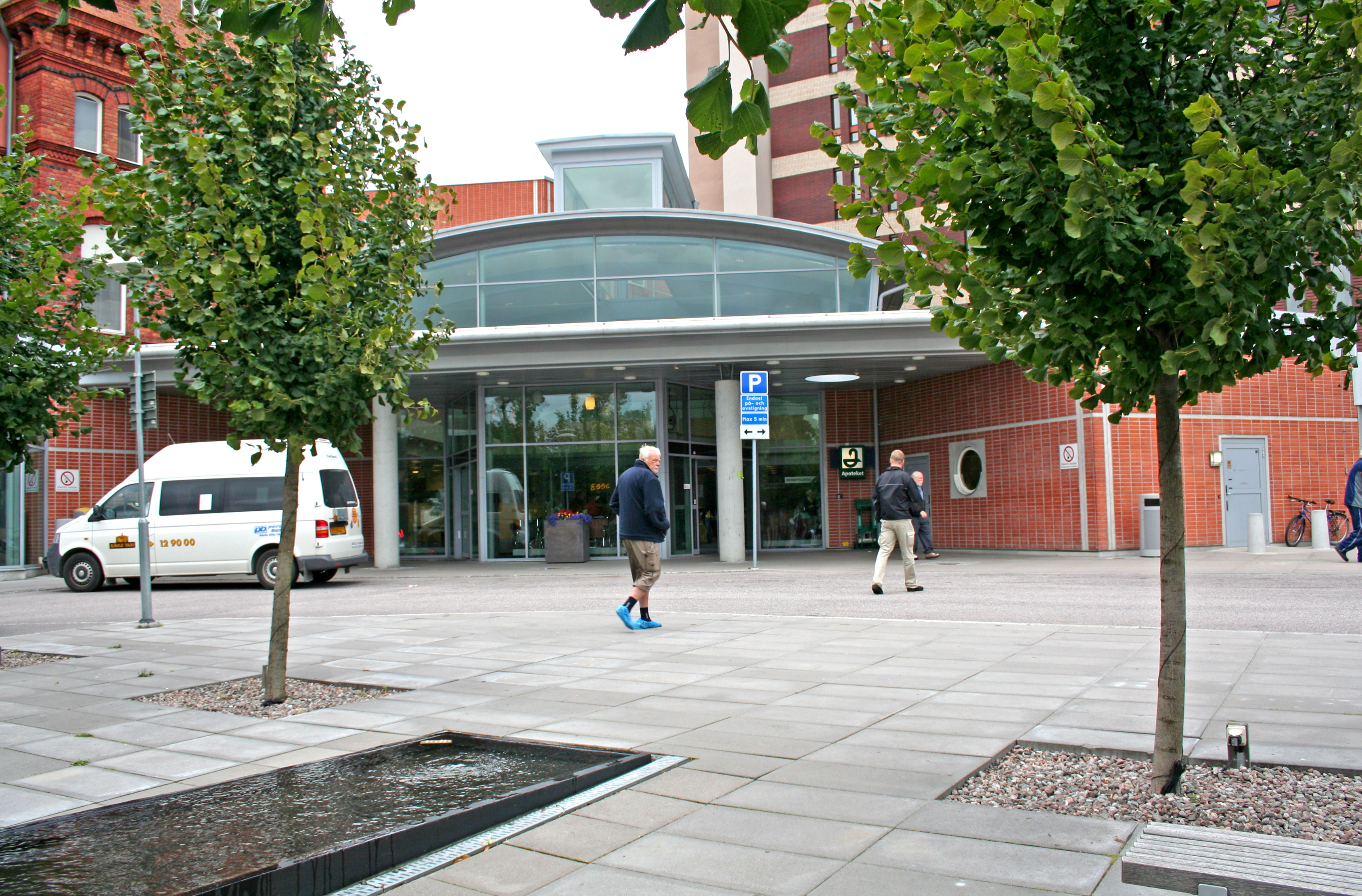
 Gävle Hospital
More efficient multidisciplinary team meetings, a greater opportunity to consult specialists at other hospitals, and a promising potential to allocate the workload during peaks. These are some of the advantages of a digitized pathology lab pointed out by Iréne Silverlo, a cytotechnologist and head of Gävle Hospital’s unit for clinical pathology and cytology. After several years’ experience with a digital workflow, she also has exciting ideas about how to gain even more benefits from digital technology.
I only see advantages to digitization. Better patient safety thanks to a reduced risk of mixing up patients and tissue samples, greater ease in comparing new and previous examinations, and better ergonomics are only some of the benefits.
Gävle Hospital has one of the first digital pathology labs in Sweden. Along with several other county councils, they are participating in a large digital pathology research project whose objectives include improving Swedish cancer care. They have been using Sectra’s digital pathology solution since the autumn of 2014.
Today six pathologists work at Gävle: two with specialist training (ST) and four pathology specialists. Two of the specialists perform their reviews essentially completely digitally, and the other pathologists also review most of their examinations using Sectra’s pathology workstation.
“I only see advantages to digitization,” Silverlo says. “Better patient safety thanks to a reduced risk of mixing up patients and tissue samples, greater ease in comparing new and previous examinations, and better ergonomics are only some of the benefits. We have also become a more attractive workplace because we are way out in the forefront of technology and we can also offer the flexibility to work remotely. One pathologist associated with us works from home—an attractive option for a specialist who is in demand.”
Gordan Maras is one of the ST doctors who uses Sectra’s solution at the Gävle pathology lab.
“We have more and more complex cancer cases to manage, and digitization has meant that it’s a lot easier for me to consult my colleague who works remotely, for example,” Maras says. “This really allows us to cut down response times.”
Moreover, Silverlo sees potential for the future in being able to share specialist expertise with other hospitals in the region or even nationwide, and to do so globally in the longer term. Digital images can be shared and discussed in real time with specialists, regardless of the geographic distance. Digitization could even mean improvements in issues such as work peaks.
“We’ve taken a look at how our radiology department cooperates with radiologists who review images remotely from other parts of the world such as Australia—an amazing opportunity to cut down response times by utilizing all of the hours of the day. We live in a reality where there’s a serious shortage of pathologists, and with digitization you don’t need to send slides around, which opens up completely new opportunities to allocate work when there are peaks.” Silverlo concluded by pointing out yet another advantage that digital technology could bring about.
“Training ST doctors is a huge challenge right now. A lot of the time we can’t take care of them as well as we would like due to the shortage of pathologists,” Silverlo says. “In a digital environment, one doctor can teach many people remotely using such technologies as video conferencing.”
However, Silverlo points out that the advantages for the reviewing pathologists are completely dependent on the digitization of the actual specimen having been done properly. A correctly prepared specimen is absolutely decisive to the quality of the digital image.
“One common misconception is that all you have to do is start scanning. But once you’ve decided to digitize your tissue samples, one of the first steps is to adapt your procedures and the process of preparing the specimens,” Silverlo says. “It might be a matter of how you put the specimen onto the slide or covering it with a coverslip or plastic film. We’ve learned a lot on our digital journey.”
“The workstation has to be user-friendly—user-friendliness is all-important,” said Maras. “I’ve tested several different workstations, and Sectra was the easiest to use. The system also has to be stable—we don’t have time for it to crash.”
Gordan Maras also sees major advantages to how he can work with images now.
“Measurements, text annotations, calculations and notes can be performed in a completely different way thanks to Sectra’s solution. It saves time and cuts down on errors due to the human factor, which increases the quality of the review,” Maras says. “Moreover, tumor diagnosis is made more efficient since it becomes more objective and exact.”
“Digital images also give you a completely different overview, compared with looking at an image through a microscope,” Maras continues. “You see the entire image, and you can zoom in on specific areas.”
“The ergonomics are also much better in a digital environment, where the stresses on your back and neck are reduced compared with your position when you work at a microscope,” he concludes.
More efficient multidisciplinary team meetings are yet another major benefit that the department has experienced from digitization. Every day at least one round is held at the Gävle pathology lab, with different categories of doctors such as surgeons, radiologists, oncologists and pathologists attending.
With digital images the secretaries don’t have to take out the slides, which used to a lot of time, and it’s easier for the pathologists to prepare the images for the rounds, which in turn makes the rounds clearer and more time-efficient.
“Preparing for the round goes much more smoothly now,” Maras said. “Since we can add comments and annotations to the digital images, showing changes or important discoveries in the image is easier, for example. This makes it easier for clinics to understand our interpretation of the image and the reason we pathologists drew the conclusion that we did. Moreover, we spend our time even more efficiently now, since before digitization it could take extra time to hunt for the discoveries you wanted to show on the slide in the microscope, for example. Since we can prepare the image digitally before the round now, we can show the discovery right away instead,” Maras concludes.
