Region Östergötland

Digital pathology—enabling pathologists to keep working efficiently during the COVID-19 pandemic

The use of digital pathology is steadily increasing and has helped the early adopters within the field of pathology globally to keep a safe social distance during the ongoing coronavirus pandemic. We talked to Mats Wolving, an experienced GI Pathologist at Linköping University Hospital, to hear his thoughts on digital pathology and how he—thanks to this technology—has not had to make any further investments or workflow changes to maintain efficient pathology operations during this worldwide pandemic.

Sectra: How long have you worked with digital pathology, and is your work situation different now compared with before the coronavirus outbreak?

Mats: I have worked full time with digital pathology since September 2019, which is when I started my position in Linköping. I have worked for many years in “regular” pathology, but I have always had a great interest in digital pathology. One reason for coming to Linköping is the fact that the university hospital here has been world leading in the field of digital pathology for a decade.

My work situation today looks very much like before the pandemic, except for the fact that many physical meetings are not taking place anymore. I alternate between working from Linköping and Gothenburg, where I live, and thanks to the technology I can do the majority of my job from any location.

In your opinion, what are the biggest advantages of working digitally?

I think this way of working offer a lot of possibilities; the first that comes to mind is how easy it is to access cases independent of your physical location. We used to be dependent of physical objects; the glass slides, microscope, and physical archive. Now, this is all accessible from anywhere and not tied to a specific location. Other opportunities are uncovered when you shift from looking in microscopes to performing the review on digital images. The digital images are scanned and instantly uploaded to the digital pathology system, part of a central enterprise imaging system, and can then be shared with other pathologists—it is easy to collaborate and consult each other without having to be in the same room. Furthermore, the added functionality that comes with being able to perform exact measurements as well as image analysis means a lot. As does the future perspective of AI assistance for both smarter workflow management and decision support.

To sum up, I would say digitization has made our job more fun and faster as well as more efficient and accurate. Digitization provides an opportunity to assure the quality of our work in areas where it has previously been a challenge to make objective evaluations. By working digitally and gaining access to more precise and automated tools, we have been able to increase the standardization of our evaluations. This is paramount in order to provide accurate and equal treatment of patients.

How has the coronavirus pandemic affected your department’s way of working?

One immediate change that we made was to cancel most of our physical meetings. Now we use virtual ways of communication, for example, the built-in chat function in the diagnostic workstation. Everything from department meetings to multidisciplinary team meetings (MDTs) are held virtually. Another way to communicate with my residents is through the annotation function in Sectra’s application, where you can highlight things and make notes for each other to see. Before the pandemic, it was more common to sit together and look at cases in front of a microscope. Another temporary impact of the current situation is that there is a decline in elective cases since fewer people are being called in for routine examinations and procedures.
To sum up, I would say digitization has made our job more fun and faster as well as more efficient and accurate.

Mats Wolving, GI Pathologist, Linköping University Hospital

You mentioned the annotation function, can you tell us more about how you use that?

I use it to make notes, both for myself and for others to see. For example, when we worked with glass slides in the microscope, it was common to write on the slide with overhead markers. Now when we are using digital images, I can make arrows, mark areas, and make notes more accurately directly on the digital image. This kind of asynchronous communication works especially well when I work with my residents—they can add questions and comments that I later review and reply to in the same annotation. It is a good way of giving feedback and we can have discussions on a more detailed level. I think much information risks getting lost and discussions tend to become less in-depth, and more on a general level, when cases are discussed together while looking into a microscope. In addition, the way of working and teaching digitally ensures that no educational opportunities are lost due to long distances, for example, if I am in Gothenburg one week and the resident is in Linköping.

Sectra recently released a new, updated view-sharing function in the application, is that something you use when you are training new doctors?

I think it might be a big perk in the future, to be able to educate in real-time from another location or for live second opinions. I can also see how it could be useful when more than two people are working together and have made separate annotations in the case; it is less of a hassle to use the view sharing than having to sit close together to do the job.

Is there any way to measure how much more efficiently you work now, compared with when everything was analogue?

As far as I know, there has not been any official measurement of that sort. What I can say is that I can see major gains in this way of working for pathologists, technologists and administrators. Since we do not have to carry glass around, all cases are in one place and everything can be compared on screen—and this is a huge advantage. In my area of expertise, we frequently want to look at old cases to compare these with more recent ones, for example, to see the progress of certain inflammatory diseases or different stages of cancer, which in the past resulted in many trips to the physical archive. Now it is all gathered in the digital archive, and the quality of older cases does not deteriorate over time. I would say there are huge logistical gains in working digitally with pathology.

When working from home, I experience that the diagnostic application from Sectra actually works better than other software systems I rely on.

Mats Wolving, GI Pathologist, Linköping University Hospital

One last question. Many readers are probably curious about your workstation when you review from home, could you describe it?

It is not as flashy as my workstation at the hospital, but it is still sufficient to do a good job. I am now trialing a 4K monitor that has a great panel and brightness, but not as finely calibrated as a radiology display, for instance. Since we work more with finding patterns and objects’ distance to each other, we don’t need the ability to see exact grayscales or shades of color.

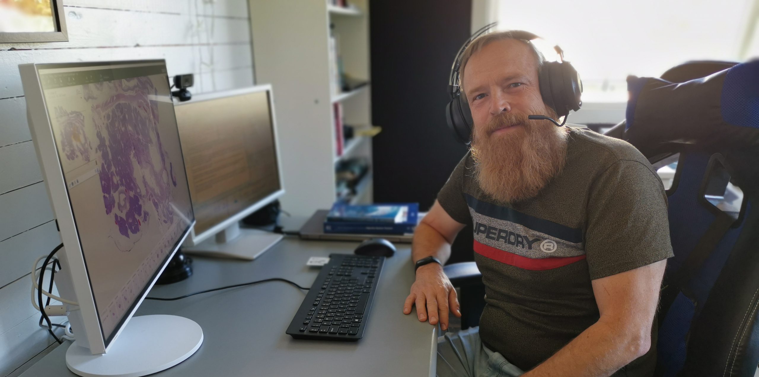
I have a mobile workstation according to the Sectra specifications equipped with, for example, a Quadro P 1000 graphics card. I use two monitors to get a good overview, and I also have a wireless headset for dictation.

When working from home, I experience that the diagnostic application from Sectra actually works better than other software systems I rely on. If I am waiting for lab data, for instance, other systems are usually really slow and keep loading, but Sectra’s solution offers high speed. I am really happy with that, and I have talked to other pathologists who have the same positive experience with Sectra’s system and the speed of loading images.

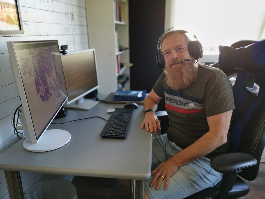
Mats in front of his workstation at home.

Featured products & services