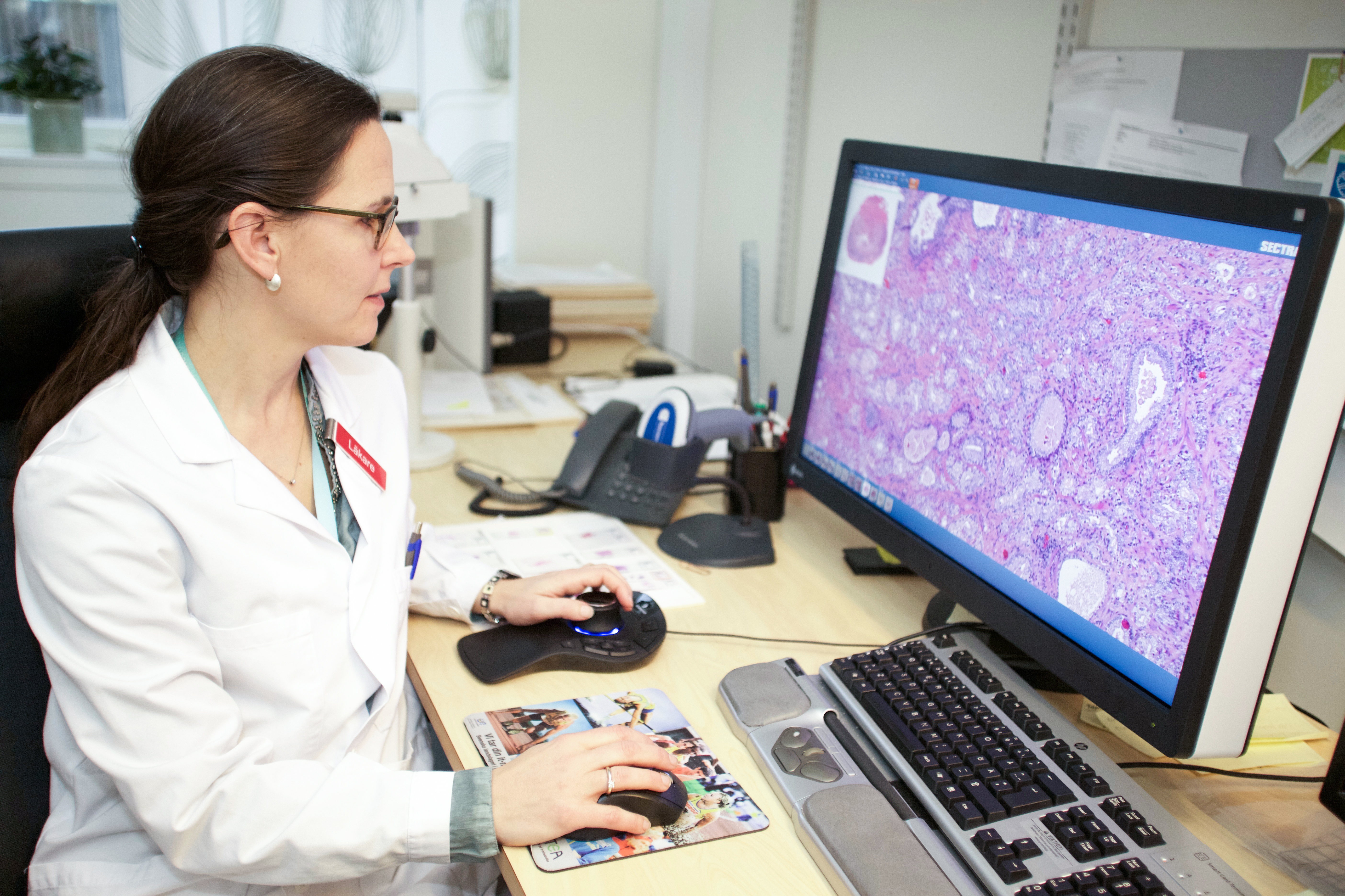
Since 2012, Linköping University Hospital has been at the forefront of medical imaging and part of a large Swedish innovation project tasked with exploring the potential digitization of pathology. The pathology department in Linköping employs about 20 pathologists, has four whole slide imaging scanners from two different vendors connected to Sectra pathology PACS and handles approximately 30,000 histopathological requests per year. All of the histopathological slides, about 180,000 slides per year, are scanned and stored digitally. As of March 2016, the archive contains a total of 760,000 scanned slides, amassing the largest clinical collection of digital pathology slides in the world. Today, consultant histopathologist Anna Bodén, who has been a key player in the project, shares the insights she has gained from working on cases in a digital environment.
Sectra’s solution is fast, easy to use and provides constant availability. You can tell that it has been developed in close collaboration with pathologists.
Tools for more efficient reading
“I read and report all my cases digitally and I feel very confident. There’s no doubt it has increased efficiency. Earlier, availability of each slide was limited to physical access only. Digitally, there is now access to the image independent of the physical slide, which is a major advantage,” says Dr. Bodén.
Dr. Bodén also points to the advantages offered by digital tools, such as being able to quickly change the orientation of an image, take measurements and make annotations. She also highlights the quick patient overview provided. In cases where she could previously see that a patient history existed, she can now—with a click of the mouse—open the relevant images together with the patient history, which helps when making her diagnosis.
“Not only does the digital solution make it easier to measure areas, etc., it also makes it much easier to find my way back to the spot where I left off, if for some reason I’m interrupted during the 30–40 minutes it can take to read a complex case. This is something I appreciate.”
Other benefits that Dr. Bodén points out are that, as the department can now retrieve all cases digitally, the need to search for glass slides is eliminated, while the ability to rapidly review earlier slides improves quality.
Availability and performance
During autumn 2015, Linköping University Hospital completed their broad roll-out of Sectra’s digital pathology solution in its pathology department. The twenty or so pathologists working at the department have now access to state-of-the-art digital pathology scanning and viewing systems, but are still able to choose whether they wish to read in the microscope, on-screen or using a combination of the two. To help them see the benefits of digital reading, Dr. Bodén points out that system performance, stability, usability and the quality of the scanned tissue are key. To allow transition from microscopy to digital, it is essential that the digital system has at least the same level of system performance in terms of availability and speed in interacting with the image as the microscope.
“Sectra’s solution is fast, easy to use and provides constant availability. You can tell that it has been developed in close collaboration with pathologists,” says Dr. Bodén.
IT department—a key role in successful implementation
Another recommendation from Dr. Bodén concerns the collaboration with the local IT department. At Linköping, the two departments work very closely together and both confirm the positive impact that this collaboration has had on the project.
“We need to provide an IT environment that meets the clinical requirements of the pathologist. I believe that working extremely closely together is a key success factor,” says Carl Clasén, Medical Technical Engineer at the hospital’s IT department.
Enhanced MDT meetings
Going digital has also dramatically increased efficiency when preparing for MDT meetings and improved flexibility during the actual meetings. At Linköping, MDT meetings for breast cancer are held once a week and usually cover about 20–30 patients per meeting.
Previously, a secretary would prepare for the meeting by collecting all of the glass slides and also print paper copies of all relevant patient information required for a particular meeting. The pathologist would then go through all of these slides and pick out the slides he or she considered to be relevant which would then be displayed using a microscope at the meeting. Today, the pathologist can easily prepare for the meeting digitally, which dramatically reduces the workload of the secretary and makes preparation easier for the pathologist. He or she can quickly go through the cases, see any notes on the images and decide which images to show. Displaying the images digitally during the meeting also offers a clinical advantage because the quality of the image is enhanced for the other participants in the MDT meeting.
“We had a big ‘Wow!’ moment in the MDT group when we started displaying our images digitally,” Dr. Bodén recalls.
Linköping also hosts regional MDT meetings. Dr Bodén hopes that their digitization will bring the possibility of sending and sharing cases digitally between care givers in the region. That would eliminate the dependence on glass slides and increase efficiency in these meetings as well.
Other related aspects include consultations and workload balancing. These are also areas where Dr. Bodén sees major opportunities in terms of increased efficiency thanks to a digital environment. However, this is not on the immediate agenda for Linköping University Hospital.
About the digitization at Linköping
Linköping University Hospital began its digital journey already in 2010 under the leadership of Sten Thorstenson, then a Senior Consulting Pathologist at Linköping University Hospital. Read the full scientific article, “Implementation of large-scale routine diagnostics using whole slide imaging in Sweden: Digital Pathology experiences 2006–2013,” in Journal of Pathology Informatics 2014, Volume 5, Issue 1, Page 14 for more in-depth background information on where Linköping stands today.
Featured products & services
Related cases


