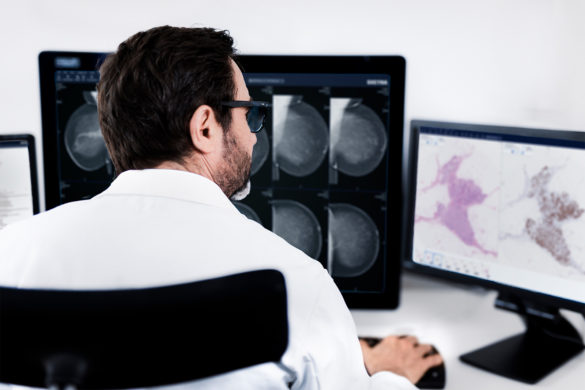
Introduction to the DICOM standard for digital pathology and its importance for workflow efficiency
White papers | Digital pathology

Industry reflection
By Simon Häger, Product Manager Sectra Digital Pathology Solution
I am confident that the advantages of digital pathology outweigh those of the analog workflow using a microscope. However, the benefit of glass slides being ‘vendor neutral’ in terms of being reviewed with any brand of microscope has not yet been realized between existing file formats and digital pathology systems. For digital pathology to become mainstream, it is crucial that scanners and viewers provide the same level of interoperability.
There is currently limited compatibility between the various digital image file formats and image viewers. Historically, pathology departments only needed a solution for reviewing low volumes using a single scanner, mainly for teaching and education purposes. Since no technical guidance existed back then, such as the DICOM standard for digital pathology, vendors optimized their file formats for fast scanning and working together with their own viewer software.
Today, many pathology departments are aiming for full digitization and are moving to enterprise solutions, demanding several different scanners to be connected to one image management system and images to be reviewed from one selected viewer. There is also a growing demand for pathologists to be able to view images from other disciplines, such as radiology and nuclear medicine. All in all, the development we are seeing now in digital pathology is increasing the requirements on image management systems and viewers to be able to consume different file formats.
Positively, some scanner vendors are responding to their customers’ requirements by providing “toolboxes” (software development kits, SDKs) and integration specifications to other vendors to enable the consumption of their files. Unfortunately, other vendors are still providing closed so called “end-to-end” systems, where the pathologists have no other option than to review slides with a viewer of the same brand as their scanner.
I claim that the “end-to-end” approach significantly hinders a full utilization of digital pathology, obstructing gains for patients in the form of the shorter waiting times and more accurate diagnosis and improved quality that this technology offers. Taking away the pathologists’ freedom to use any viewer and image management system in combination with any scanner makes the sharing of images with colleagues very difficult. It also increases costs as it prevents an implementation of best-of-breed scanners. “End-to-end” systems also stand in the way of the possibility to consume images from other disciplines, obstructing one of the biggest and most positive changes we see in healthcare imaging today, namely integrated diagnostics. In addition—and perhaps most importantly—it makes it difficult for the healthcare provider to implement and benefit from new innovation happening outside the scope of their “end-to-end” vendor.
The majority of pathologists I have met want to use different scanners for different purposes, but to interact with only one user interface, i.e. one viewer. In order to provide patients and referring physicians with the highest quality diagnosis with less waiting time, the pathology department needs to use the best-of-breed in scanners. Some scanners are good at high-volume production, some at frozen sections, and some vendors are experts in providing low-cost desktop scanners for telepathology purposes. You probably have some in mind under each category while reading this.
There is also an efficiency gain to be considered by using one viewer, given that it is fast, since the pathologist will learn its features well and trust what he or she sees when getting up to speed in reviewing images digitally. Ultimately, it should be up to the pathology department—and not the scanner vendor—to decide if the pathologists should review the images with a viewer from Sectra, Philips, Leica, Hamamatsu or any other brand.
As I mentioned in a previous article, the DICOM standard for digital pathology (supplement 145) is working towards a harmonization of file formats and communication protocols between different solutions to solve these kinds of integration issues. Several scanner vendors that I have talked to assume that DICOM will be the file format of the future in pathology, and therefore develop new functionality to be DICOM compliant. Although the adoption of the DICOM standard in digital pathology looks promising, it seems like it will take time before the majority of vendors comply with it.
Until the DICOM standard is in place, it is up to the buyers and users of digital pathology scanners, viewers and image management software to demand that these are open and can be integrated into one efficient digital pathology system. To reach a highly efficient system, it should be up to the buyers to “cherry pick” components for this system to be optimized for their workflow.
There are many ways to solve this interoperability problem. The proprietary whole slide image files are not a problem in themselves; they are often optimized to allow for fast scanning, which is crucial for creating an efficient digital workflow. What does become a problem is when the scanner vendor does not allow any other software than its own to consume these images. Vendors should preferably describe how the files are structured and can be read by other applications, and provide SDKs for reading the images.
The trend is positive, and more and more vendors have understood the benefits of providing open interfaces and the drawbacks of “end-to-end” systems. Those closed systems that still won’t integrate with other vendors argue that this is for safeguarding the quality of their entire system. However, this argument has proved to fall short many times in other industries, and it is probably just a way for these vendors to lock in their users to only use their applications.
Glass slides can be reviewed with any microscopes, but today digital pathology cannot offer the same interoperability between the scanners, image management software and viewers. Digital pathology must at least offer an interoperability comparable to the microscope and the slides to enable a digital transition. A broad adoption of the DICOM standard will—when fully implemented—solve this in the end. Until then, buyers of digital pathology systems need to demand interoperability and openness in order to realize the full benefits that digital pathology promises.