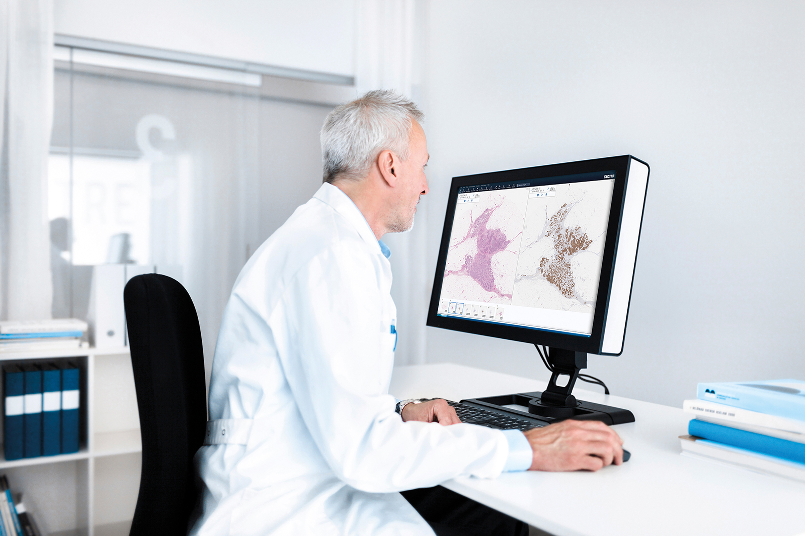
Until recently, using a conventional light microscope has been standard practice for examining histopathology slides. However, the use of digital pathology is on the rise and many pathology labs are now investing in scanners for digitizing glass slides. As pathology departments move from analog to digital workflows, many aspects of their daily work will be affected. To a large extent, these changes mirror those that occurred in radiology departments 10 to 15 years ago.
The aim of digitization is not to let the computer assume the role of the pathologist, but to use computers for time-consuming work that is difficult for the human brain to process, thereby freeing up valuable time that can be used by the pathologist to provide the correct diagnosis. Other advantages with the digitized workflow include the possibility for a more active production planning, allowing staff and managers to keep track of how many specimens are in the system, where in the production chain they are, whether time limits are being kept, etc. Digitized images also eliminate the need to go to the glass archive when looking for a previously prepared slide. In addition, while glass slides can be broken or lost, a digital image will always be stored in the correct place and thus be easily found.
This text aims to illustrate the fully digital workflow at a pathology department, describing the use of hardware, software and IT architecture.
The electronic referral
The pathology processes start with a referral; this is no different in a digital workflow. In a fully digital workflow, the referring department issues an electronic referral through integration between the electronic health record system (EHR) at the referring department and the information system at the pathology department. The EHR and the request system provide input data about the patient, specimen, medical history and reason for referral. Integration between the two systems reduces the risk of specimen mix-ups and thus increases patient security. It also saves time compared to manually entering data into the information system used in the lab, the laboratory information (management) system, LIS or LIMS for short. A LIMS could be described as a more modern LIS, which can also manage workflows and guide the secretary, technician and pathologist through the process with checkpoints for the different steps in the lab.
Arrival at the pathology lab
When the specimen arrives at the pathology lab, it should preferably have been marked by the referring department with a barcode, which links the specimen to the correct referral. At the pathology department, the barcode is scanned and thereby registered in the LIMS as having “arrived”.
Preparation of specimens
Many steps in the specimen preparation process are performed manually, and this is also the case in the digital workflow. However, in the non-digital workflow, it is often impossible to track where specimens are in the production chain. By digitizing the process, using barcodes and “checking in” the specimens and slides at the various stations in the lab, it is easier to identify and learn to avoid bottlenecks, to see whether time limits are being kept, etc.
In the following sections we will describe some of the hardware and software involved in the preparation of specimens.
Digital macro cameras: Digital macro cameras are used to capture images at the macro grossing station. These images aid the pathologist in orientating the origin of each slide and eliminate the need to sketch a macro image manually. Integration of the macro camera into the LIMS and digital archive ensures that the correct macro image is shown together with a particular slide, again reducing the amount of manual work and the risk of case mix-ups.
Scanners: Investing in scanners is often the first step in digitizing a pathology lab. Depending on your needs, there are different options available in terms of capacity, level of automation, etc. Scanners with a capacity to load several hundred slides and provide whole-slide images (WSI) in different file formats are available on the market. Scanning glass slides creates very large files and it is therefore important to decide what information is really needed, since this influences the choice of magnifications and focus planes. At the same time, for diagnostic purposes, the digital image must be at least the same quality as the glass slide viewed in the light microscope.
To minimize the number of re-scans, there is a higher demand on section quality compared with the preparation of glass slides for light microscopy. A thin section generates higher-quality images than a thick section, since it facilitates the identification of the correct focus plane, thereby speeding up capture times [1]. Furthermore, since it can be difficult for scanners to scan the edges of a slide, it is preferable that sections be placed in the middle of each glass. This enables the scanner to better identify the borders of the section.
Use of barcodes: Many scanners include a 1D or 2D barcode reader, which can help integrate the scanner with the local LIMS. From a patient-safety perspective 2D barcodes are preferred, as these contain a checksum ensuring that a barcode that is read is done so correctly. A 1D barcode does not include this built-in control.
The barcode links the slides to the correct specimen, and can, for example, include information about applied staining and scanning protocols, such as whether one or several focus planes should be applied and whether a x20 or x40 magnification is needed.
One thing to keep in mind is that certain scanners, staining apparatus or other hardware, are equipped with readers that are only able to read specific types of barcode (1D or 2D, for example). This may implicate having to re-label the slides, for example, between immunohistochemical staining and scanning. However, there are barcode readers available on the market that are compatible with various barcode-types. Thus, by making careful choices in configuration of barcodes, it is possible to avoid the need to re-label slides, something that not only takes time but also increases the risk of mix-ups.
Magnification settings: There is an ongoing discussion as to whether a x20 magnification is sufficient for the purpose of reviewing or if images need to be scanned using a x40 magnification. Doubling the magnification increases the size of the image by a factor of four and, in older scanners, can prolong the scanning time by a factor of about three. However, newer scanners take only a marginally longer time to scan in x40 magnification compared to x20 magnification. Validation studies of the use of WSI for diagnosis using a x20 magnification have shown a concordance with glass slides of over 80% and in many cases over 90% [2-8], indicating that a x20 magnification is sufficient for clinical diagnosis in most cases, while x40 can be applied when needed, for example for mitosis counting.
Focus plane/s: When reviewing glass slides in a microscope there is often the necessity to adjust the focus due to uneven surface of the specimen. With modern scanners, locally adjusted focus points are used when scanning the specimen, creating a WSI with optimal focus over the entire section, thus eliminating the need to refocus the image continuously during review.
Many scanners also offer the possibility of scanning with multiple focus planes, useful for example in cytology, when different depths of the image need to be reviewed. This creates larger data files and should therefore only be applied when needed, rather than as a standard protocol.
The diagnostic reading
A digital workflow moves the pathologist’s primary workstation from the microscope to a computer screen. Depending on the IT solutions used by the department, it may also be possible to access the image viewer and any additional information needed from outside the hospital, for example from home.
In the following section we will describe some of the hardware and software encountered during the diagnostic reading.
The digital work station: The digital workstation offers several advantages compared to a microscope. One is the possibility of viewing several images, for example sections with different staining techniques, side-by-side for comparison. This can facilitate the interpretation of the section. Current images can also be compared against old, stored images from the same patient. Distance and area measurements and annotations can also be facilitated using digital images.
There is still a debate as to whether reviewing digital pathology images is more time-consuming than working in the microscope. However, using a digital workflow eliminates the need to find and change glass slides, match the slide with the patient, find the right focus, perform manual measurements, etc. All of these things take time, and it has been shown that when reviewing a case, 43% of the time is used for other tasks than looking at the actual slide [10].
With a basic viewer, that previously dominated the market, a slow-down in the actual review speed was noted. However, it has been shown that by using a modern, well-functioning viewer, it is possible to perform the review as quickly digitally as in the microscope, despite having limited training in the digital system [11].
Computer screens: Ergonomics is an important driver of the digitization of pathology, and while working too long in front of a computer can also cause problems, this way of working does not impose the same static position of looking at sections through a microscope. For an ergonomic and convenient workflow, it is recommended that the pathologist has at least two computer screens at his or her review workstation: one for monitoring patient and specimen information typically found in the LIMS, and the other for image review. Any general computer screen can be used for patient and specimen information, while the screen used for diagnostic review should preferably be a larger and with higher resolution to make it convenient to review the sample. The variations originating from staining and scanning are larger than the variations from a regular calibrated screen [9], suggesting that a medical grade screen is not needed. A 27” screen with 4 megapixel resolution, or 30” with 6-8 MP resolution, is often appreciated by the reviewer as well as a high refresh rate.
Image analysis software: With the addition of image analysis software, it is possible to receive assistance, for instance in the quantification of immunohistochemical staining and mitotic counting. FDA approved tools are available for automatic quantification of, for example, the number of Ki-67 or HER2 cells [12-13]. This saves time for the pathologist and can increase reproducibility. As no algorithm is perfect, allowing the pathologists to confirm or correct the result is often preferred.
Reporting: Depending on the specific workflow and system set-up, reporting can be carried out in the LIMS system or in the image viewer. Again, integration between the reporting system and the EHR is advantageous, since this reduces the need for manual work when reporting back to the referring physician.
IT structure
Digitizing histopathology slides will generate large sets of data, creating a need for adapted storage solutions. Each glass slide generates a file of several hundred MB, and in some cases several GB. The digital images can be stored locally at each hospital, or centrally in a particular region or country. Regardless of storage model chosen, it is imperative that the system complies with any access regulations and restrictions in accordance with patient privacy legislation and caregiver policies.
A centralized, for example cloud-based, storage system offers several advantages. A central storage system can be organized in a more cost-effective way due to both the large volumes and the varying demands imposed in terms of uploading and downloading times. This type of storage solution also offers the possibility of shared support and maintenance solutions, as well as system administration. Updates to the system can be rolled out centrally instead of locally, thereby reducing deployment costs. It would also be possible to have a certain degree of central integration with other systems, such as image analysis software.
By applying image lifecycle management rules on the storage, one can ensure that images are stored in the right place (using, for example, a tiered storage solution) or that only parts of the images are stored long term.
The bandwidth needed to enable pathologists to work at an acceptable speed depends on the amount of uploaded and downloaded data, as well as the number of users logged into the system at any given time. Bandwidth between scanner and storage solution is more critical than between image storage and reading client. Uploaded data includes the number of scanned glass slides at any given time, and downloaded data refers the number of images downloaded for reading at any given time. If the department’s bandwidth is very low or expensive, the user may prefer an in-house storage solution, since a centralized option could become too slow or expensive.
As review becomes digital, uptime of the system will be critical. For this reason, the IT provider should be used to large installations and the installed system should be redundant ensuring that review can always continue.
External consultation
Digital slides can be distributed for external consultation in a more convenient way than glass slides. They easily can either be sent to the external consultant, or the consultant can be given access to the system to review the images remotely. Due to image size, streaming images is preferred compared to uploading the full image.
MDT meetings, research, quality and education
WSI makes it possible to prepare and implement multi-disciplinary team meetings in a much more efficient manner than using glass slides [14].
Using structured requests and reporting enables data to be inserted into research and quality registers in an uncomplicated way. With a common system for tags, file formats, etc., a quality database can be collected at the national or international level with only a few simple steps, thus helping to maintain or increase the knowledge within the pathology community, as well as control the quality of the service at different departments. This enables an improvement in education quality and standardized training, since digital images can easily be shared between departments [Reviewed in 15].
Summary
Digital pathology is still in its infancy but is already providing tools that can facilitate the pathologist’s daily work. The possibility of reviewing images side-by-side or performing measurements in digital images are only two of the advantages offered by digital pathology. Digitizing the pathology workflow also offers an excellent opportunity to use digital tools to speed up otherwise time-consuming tasks, such as cell counting or organization of samples, thereby freeing up time for the pathologist to set a diagnosis.
References
- Yagi Y and Gilbertson JR. A relationship between slide quality and image quality in whole slide imaging (WSI). Diagn Pathol. 2013; 3 (Suppl 1): S12.
- Al-Janabi S, Huisman A, Nap M, Clarijs R and van Diest PJ. Whole slide images as a platform for initial diagnostics in histopathology in a medium-sized routine laboratory. J Clin Pathol. 2012 Dec; 65 (12): 1107-11.
- Al-Janabi S, Huisman A, Nikkels PGJ, ten Kate FJW and van Diest PJ. Whole slide images for primary diagnostics of paediatric pathology specimens: a feasibility study. J Clin Pathol. 2013; 66 (3): 218-23.
- Al-Janabi S, Huisman A, Willems SM and Van Diest PJ. Digital slide images for primary diagnostics in breast pathology: a feasibility study. Hum Pathol. 2012; 43 (12): 2318-25.
- Al-Janabi S, Huisman A, Vink A, Leguit RJ, Offerhaus GJA, ten Kate FJW and van Diest PJ. Whole slide images for primary diagnostics in dermatopathology: a feasibility study. J Clin Pathol. 2012; 65 (2): 152-8.
- Al-Janabi S, Huisman A, Vink A, Leguit RJ, Offerhaus GJA, ten Kate FJW and van Diest PJ. Whole slide images for primary diagnostics of gastrointestinal tract pathology: a feasibility study. Hum Pathol. 2012; 43 (5): 702-7.
- Nielsen PS, Lindebjerg J, Rasmussen J, Starklint H, Waldstrøm M and Nielsen B. Virtual microscopy: an evaluation of its validity and diagnostic performance in routine histologic diagnosis of skin tumors. Hum Pathol. 2010; 41 (12): 1770-6
- Wilbur DC, Madi K, Colvin RB, Duncan LM and Faquin WC. Whole-Slide Imaging Digital Pathology as a Platform for Teleconsultation. A Pilot Study Using Paired Subspecialist Correlations. Arch Pathol Lab Med. 2009; 133 (22): 1949-1954.
- Treanor D, Oral presentation, Pathology Visions, San Antonio TX, 2013.
- Randell R, Ruddle RA, Quirke P, Thomas RG and Treanor D. Working at the microscope: analysis of the activities involved in diagnostic pathology. Histopathol. 2012; 60 (3): 504-10.
- Randell R, Ruddle RA, Mello-Thoms C, Thomas RG, Quirke P and Treanor D. Virtual reality microscope versus conventional microscope regarding time to diagnosis: an experimental study. Histopathology. 2013; 62 (2): 351-8.
- http://www.accessdata.fda.gov/cdrh_docs/pdf7/k071128.pdf
- http://www.accessdata.fda.gov/cdrh_docs/pdf11/k111543.pdf
- Heffner S. Streamlining Tumor Board Reviews. Perspectives in Pathology. 2008; 17 (7): 20.
- Pantanowitz L, Szymas J, Yagi Y and Wilbur D. Whole slide imaging for educational purposes. J Pathol Inform. 2012; 3: 46.

