Digital magazine: This is enterprise imaging
Articles | AI in medical imaging | Breast imaging | Cardiology imaging | Digital pathology | Enterprise imaging | Enterprise platform | Ophthalmology imaging | Radiology imaging
White paper
There is an increasing emphasis on adding quantitative data to imaging. This improves not only individual patient care but also reproducibility and enables general healthcare outcomes to be measured. In orthopaedics, there are radiological measurements that are important for accurate diagnosis and treatment planning and should always be included in radiology reports.
In their study, Leide et al. (2021) commented that these measurements are not always taken into consideration due to limited resources. Hence, time-saving software can lead to more measurements being performed and therefore result in more patients being correctly diagnosed. An earlier diagnosis has also been shown to improve clinical outcome (Kennedy et al. 2017). Recently, easy-to-use guides using advanced image analysis have proven to be one such tool. These guides show promise when it comes to improved workflow, consistency, and fewer clinical errors. This study was performed to measure the time savings that can be gained by using one of the new guided measuring tools for hip dysplasia.
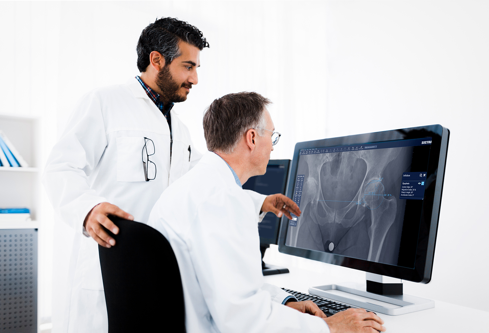
Measurements were performed by a single observer, a musculoskeletal radiologist, using a hip dysplasia guide in the pre-operative planning software from Sectra, followed by traditional manual measurement methods on AP radiographs of the pelvis in 65 subjects (130 hip joints). The lateral center edge angle, Sharp’s angle, acetabular index angle and migration index were measured both using the guide and manually, and the time needed for each method was recorded. A table with the measurements was created automatically by the software only when using the guide. The extra time required to type the manual measurements was recorded.
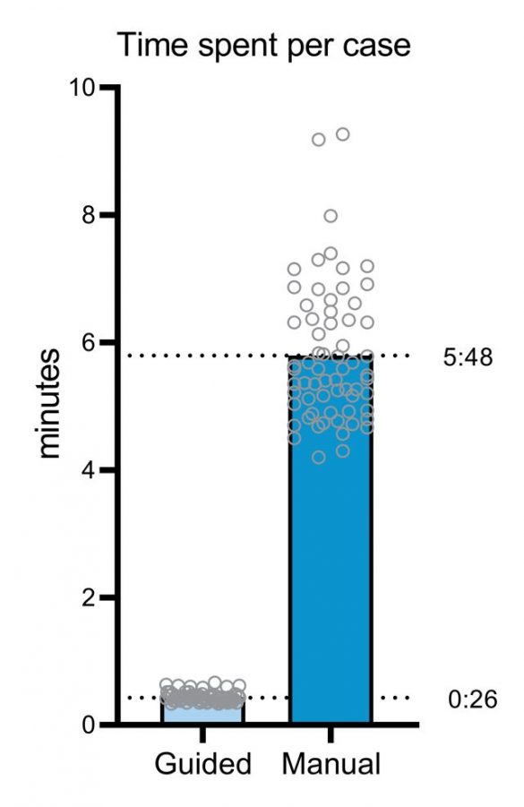
The average time for the measurements using the guide per patient (two hip joints) was 26 seconds (range 20–40 seconds). The average time for the manual measurements per patient (two hip joints) was 222 seconds (range 164–296 seconds). The manual typing of the measurements took an average of 121 seconds.
Based on the time savings recorded in this study, a hospital performing 500 hip dysplasia measurements per year can typically save a work week, approximately 40 hours, per year. In addition to greatly increased time efficiency and therefore work throughput, these tools can also help shorten the learning curve for less experienced radiologists since they eliminate the need to research how peers perform the measurements and which landmarks to use. Automatic calculations and the ability to easily copy and paste data to the RIS/HIS could potentially minimize the risk of human errors.
Significant time savings and increased efficiency could be achieved by using these guided tools for image analysis and measurements. Hence, integration of the tools into daily practice shows promise when it comes to improved workflow and standardization of care.
Rebecka Leide, Anna Bohman, Daniel Wenger, Søren Overgaard, Carl Johan Tiderius & Cecilia Rogmark (2021) Hip dysplasia is not uncommon but frequently overlooked: a cross-sectional study based on radiographic examination of 1,870 adults, Acta Ortho., 92:5, 575-580
John W Kennedy, Alistair S Brydone, Dominic Rm Meek & Sanjeev R Patil (2017) Delays in diagnosis are associated with poorer outcomes in adult hip dysplasia, Scott Med J., 62(3), 96-100