Helsingborg, Sweden

Integrated diagnostics in practice: a pilot test of a globally unique solution for the investigation of prostate cancer

A working group from the National Prostate Cancer Register of Sweden (NPCR) and the Regional Cancer Centers (RCC) worked with Sectra to develop a new digital solution that could become the national standard for investigations of suspected prostate cancer. In autumn 2023, Helsingborg Hospital became the first hospital in Sweden to conduct a pilot test of the globally unique solution, which provides a better overview of the patient’s investigation and outcome thanks to integrated diagnostics, structured report templates, and automated feedback loops and can be used as a decision support tool when determining treatment.
   
With a clear vision of “a coherent flow” between radiologists, urologists, and pathologists, this will lead to better diagnostics, fewer healthcare injuries, and better patient treatment. The solution is also designed to reduce the administrative burden and increase the quality of reporting to national quality registers via direct transfer from the report templates. Now the working group is sharing the specific effects and results indicated by the evaluation of the pilot project.

All of the radiologists feel that the structured work method saved them time—from five to seven minutes per case on average, which is a lot. Say that it takes 15 minutes to review a prostate case. If five minutes can be saved per case, then we can free up a tremendous amount of radiology time every year.

Erik Thimansson, Senior Physician and PhD candidate in radiology at Helsingborg Hospital

A vision of improved prostate cancer diagnostics

According to statistics from the Swedish National Board of Health and Welfare cancer registry (Swedish Cancer Society 2023), prostate cancer is the most common form of cancer in Sweden in terms of the number of people who develop it. Every year, approximately 12,000 men develop prostate cancer, which is the form of cancer that causes the most deaths among men. However, the disease can be cured if it is discovered at an early, localized stage. PSA testing has reduced the prostate cancer mortality rate, but it has also resulted in certain men being treated unnecessarily, which causes both unnecessary suffering for patients and unnecessarily high healthcare costs.

A major breakthrough in prostate cancer diagnostics came several years ago when magnetic resonance imaging (MRI) of the prostate was introduced in healthcare. These refined MR diagnostics make it possible to identify areas where aggressive prostate cancer is suspected, which can then be tested more specifically using targeted biopsies. This has revolutionized diagnostics and become the standard in the initial investigation phase for suspected prostate cancer, but it has also brought new problems. MR images are complex and difficult to interpret, even for experienced radiologists. Moreover, it has not been possible for radiologists to receive feedback on their assessments in a straightforward and effective manner—where were the biopsies taken and what did they show?

Erik Thimansson, Helsingborgs lasarett

Erik Thimansson, Senior Physician and PhD candidate in radiology at Helsingborg Hospital

Erik Thimansson, senior physician and PhD candidate in radiology at Helsingborg Hospital, describes this as the background of the pilot project to test a new digital solution for investigations of suspected prostate cancer that they recently concluded and evaluated.

“The entire project originated with the NPCR, which has been driving the issue of structured diagnostic templates for radiology, urology, and pathology in the INCA environment for several years,” he remarks. “I encountered the initiative for the first time in September 2020, when we were going to begin OPT, organized prostate cancer testing, in Region Skåne. All 50-year-old men in the area had the option to participate in OPT, which included taking a PSA test and, in the event of elevated PSA, an MRI prostate test. Biopsies were performed if serious cancer was suspected based on the MRI or PSA density. In connection with this, in Helsingborg, we began to use structured report templates and required all OPT cases to be registered in the INCA environment. At the beginning of 2022, we installed the template for prostate MRIs linked to INCA developed by Sectra in close partnership with Fredrik Jäderling, radiologist at Capio Saint Göran’s Hospital in Stockholm, and Magnus Törnblom, urologist at Visby Hospital, who became chairman of the national working group.”

Eric explains that the international PI-RADS standard is used when reviewing prostate cases in order to adopt a structured approach when assessing the risk of significant cancer. The report template is based on PI-RADS, and it directs the radiologist—in a positive way, he clarifies—to look at the right things in the investigation in a structured manner. “The radiologist is assisted by a combination of a structured review and a structured opinion. The template caught on quickly here at Region Skåne, not just for OPT cases but for ordinary prostate cases as well. It saved a lot of time, provided assistance, and provided a good structure for the report. Moreover, we have to localize lesions in sectors and visualize them in a biopsy template, and this template provides that ability as well, so that was a major additional benefit.”

Now opinions from the radiologist in Helsingborg that reach the urologist have the same structure every time, which Eric confirms the urologists truly appreciate. “Previously, our opinions varied a lot, with a lot of rambling text. It was frustrating for the urologists when they couldn’t quickly understand what was important. Now every opinion begins with a summary of the most important things first, and then come observations for each suspected change. And they automatically have access to the biopsy template, where they can see where the lesion is located on three different slices.”

Once they had been using the template and the automatic link to the INCA registry for several months, Erik contacted the NPCR to discuss some suggested improvements. “I showed them what we were doing in Skåne and how effective this prostate template had been for us, and explained that the link between Sectra PACS and their quality registry provided automatic registration. They were extremely interested—both in doing better work in clinical terms and in automatically registering the data in a national registry. ‘This is the future,’ they said.”

The NPCR had a long list of requests before startup, and Sectra took the list and was able to implement it. It was wonderful when we got it to work—we were practically saying ‘hallelujah’ here.

Erik Thimansson, Senior Physician and PhD candidate in radiology at Helsingborg Hospital

At the end of 2022, the RCC began to set priorities and focus on the national structured diagnostic templates for MRIs, biopsies, and pathology in prostate cancer diagnostics,

Johan Ivarsson, RCC väst

Johan Ivarsson, Product Owner and IT Project Manager at Regional Cancer Center West

and they appointed a working group to continue working on them at a faster pace. Johan Ivarsson, product owner for various solutions on the INCA platform and technical project manager at the RCC, was given the responsibility for leading technical development.

“At the beginning, my job was to put together the working group consisting of urologists, radiologists, and pathologists, as well as take stock of the solution in place at the time: what it looked like, what it should look like in the future, and what they wanted to achieve with the product,” Johan explains. “We agreed on a clearer vision of the purpose of the solution and who the stakeholders are. In general, what should the solution result in, and for whom? Next, we worked on compiling requirements and gathering all of the nodes that were concerned and involved, including Sectra for the MRI part, as well as INCA for the patient overview with links and data transfers for quality registries and OPT.”

The right conditions for a pilot test in Skåne

The solution that was developed is based on making all of the documentation related to MRI investigations, biopsies, and pathology reports structured and digital. Thanks to the digital structure, the information can be transferred directly to the medical record and to the prostate cancer registry if prostate cancer has been established. The working group saw several advantages with the solution in theory, but the integrated flow needed to be properly evaluated.

Region Skåne and Helsingborg Hospital came up as natural candidates for a pilot test in a clinical setting early on. For one thing, they had already been using parts of the solution, and they already had the right technical capabilities for integrated diagnostics in place. For another thing, the hospital was considered “the right size” to conduct a reasonable project.

The region has been a customer of Sectra’s for many years, and Sectra’s PACS has been used as a diagnostics application region-wide in both radiology and pathology. Before the pilot project, a close dialogue and partnership took place between the hospital in Helsingborg, the RCC and Sectra to address the working group’s requests, improve the existing functionality of the solution, and develop a function for automatic feedback from INCA that is integrated into Sectra PACS. Both Johan and Erik describe the partnership as “running extremely smoothly” and “invaluable.”

“In general, technical integrations tend to be complex and difficult, but I think things went smoothly here,” says Johan. “We had a constructive initial meeting attended by a Sectra solutions architect who immediately understood our needs and requirements and could bring them to product development. A technical concept was developed before the summer, and development took place during a few weeks in August, so we were able to install the solution in September.”

“The NPCR had a long list of requests before startup, and Sectra took the list and was able to implement it. It was wonderful when we got it to work—we were practically saying ‘hallelujah’ here,” Erik adds.

The vision of the pilot project, Johan continues, was to be able to test using the three structured report templates and the functions built into the templates in practice. “Of course we wanted to evaluate the result and what the professionals concerned—radiologists, urologists, and pathologists, as well as medical secretaries, nurses, biomedical analysts, and others who file reports in the quality registry—thought about the work method and the flow. We wanted to get the operational work method up and running properly and without too much fuss, then provide the users with a thorough introduction and develop practical work processes and procedures together.”
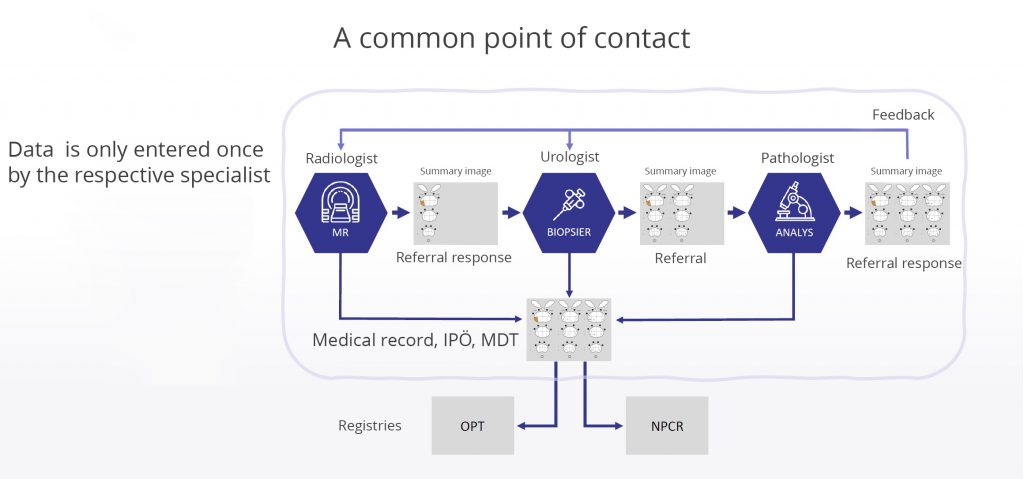
 

Overview of the diagnostic chain with a feedback loop. A single-entry model, consisting of the initial documentation by each physician, is sufficient for creating referral replies, graphic visualization in the interactive decision support tool Individual Patient Overview for prostate patients, and registration in the NPCR. Image: NPCR


The pilot test ran from early September 2023 until December 1. It included around 100 biopsy patients, and around 50 staff members at Helsingborg Hospital participated in the pilot, Erik explains. “At the hospital, I’m the person in charge of the radiology part; senior physician Viktoria Gaspar is in charge of the pathology part; and senior physician Christian Torbrand is in charge of urology. Here in Helsingborg, we have ten radiologists who write prostate MRI reports, and all of them participated. Aside from us, the pilot involved a handful of pathologists, several urologists, and several nurses in all three units that participated.”

Erik explains that the radiologists in Helsingborg, who review a handful of prostate cases per week on average, changed very little in their workflow; they were able to work as usual and submit reports using the review template in Sectra PACS, which is automatically synchronized with the quality registry. Automatic feedback from INCA was an additional feature that was activated for them during the pilot. As soon as the pathology opinion is completed, each radiologist receives feedback on their cases in an individual dynamic work list in PACS. This might seem like a small change, but it is extremely important to the radiologists, and Erik and his colleagues found it unbelievably valuable.

“It’s important to us to find out where the biopsies were taken and what they showed,” Erik says. “When we wanted to follow up on a case in the past, we either had to log in to the patient’s medical records and search, which is time-consuming, or if we were lucky, the case came up at a multidisciplinary team meeting, which does not happen for many cases. The great thing about the built-in feedback from INCA is that we now receive a summary from the urologist and pathologist directly in our diagnostic tool via a so-called web window. The feedback thus becomes individualized and automated—an unbelievably seamless solution, which is extremely valuable. Naturally, it’s good to know if I was right, but perhaps it’s even more important to find out when my assessment was incorrect. By having this feedback loop, we build on our personal experience and can calibrate ourselves and become better radiologists over time. We can also reduce what we call overdiagnosis and avoid saying that cancer is present when there actually isn’t any cancer.”

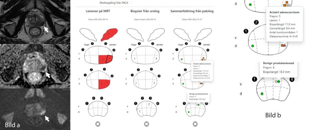
The summary view that the radiologists have access to directly in Sectra PACS. The compilation/table/summary in the Individual Patient Overview (IPÖ) is displayed via a web window. Image a) PI-RADS 5 lesion in the peripheral zone dorsolateral left side, and image display of feedback in PACS. Image b) The targeted, MRI-guided biopsies showed a Gleason score of 4+5=9 (dark red star). Some systematic biopsies bilaterally in other localizations in the prostate were benign (green dot). Images: Erik Thimansson.

A lot of time saved and overdiagnosis reduced in the long term

After the pilot was completed, an evaluation was conducted with everyone who participated in the test. The working group is now compiling the results and preparing them for publication.

“We received an extremely positive response from our ten radiology colleagues who took part in the pilot project,” Erik reveals. “All radiologists feel that the structured work method saved them time—from five to seven minutes per case on average, which is a lot. Say that it takes 15 minutes to review a prostate case. If five minutes can be saved per case, then we can free up a tremendous amount of radiology time every year.”

Erik clarifies that the time savings only relate to the use of the diagnostic template, not the feedback loop that they also have now. The feedback loop is also proving to make positive contributions on many fronts.

“For the first time, every radiologist receives feedback on their work and their performance so that they can calibrate themselves and become a clinically better radiologist over time. That’s the major benefit. If we can reduce overdiagnosis, we’ll realize savings in many areas, including financially. But the most important thing is for us to reduce patient suffering—no unnecessary tests, treatments, and checkups, which reduces both physical and mental suffering. For example, around 1 percent of biopsy patients develop sepsis, which is unbelievably serious and requires drastic treatment. A reduction in unnecessary biopsies reduces both patient suffering and the risk of complications.”

Erik also explains that everyone who was asked about the automatic feedback in the evaluation gave it top scores and made comments such as “truly excellent,” “a lot of added value,” “unbelievably valuable in every situation,” and “the only way to improve.” Keeping the information together in the same system means that it will be easier to access and take less of every radiologist’s time, so that more of them can develop professionally and improve their diagnostic skills.

“Some other things that we observed,” Johan interjects, “are that the report templates help to structure the actual registration of data, both for keeping medical records and for registration in quality registries. Just having the template is a tremendous help to the physicians, but automatic registration in quality registries has also proven to be extremely valuable—in streamlining the process and saving time, but also by ensuring that the right information is entered. We see this as a huge advantage. The administrative workload is clearly reduced, and the pilot test showed that by using the templates, it’s possible to analyze diagnostic precision and adherence to the national care program with a few keystrokes, something that was time-consuming and extremely difficult in the past.”

“We radiologists really see it as a win-win, with structured review, data sent to the quality registry, and individualized feedback in a one-stop shop,” Erik adds.

The urologist’s and pathologist’s perspectives

Urologists and pathologists also participated in the pilot project, as previously mentioned. We interviewed Christian Torbrand, and Viktoria Gaspar, senior physicians in urology and pathology, respectively, at Helsingborg Hospital. What changes did they see in their flows in this pilot project?

Before this, the reports were unstructured and hard to interpret: everyone wrote a bit off the cuff, and we had to work hard to interpret where the changes were located and how serious they were. Now the reports are completely structured instead, which helps us a lot: with our diagnoses, how to target our tissue samples and with treatment.

Christian Torbrand, Senior Physician in urology at Helsingborg Hospital

“There was very little difference for us pathologists, just as for the radiologists, since we had already been working this way for a couple of years when the pilot began,” Viktoria says. “The difference in the pilot was that the urologists also needed to enter their part into INCA, where they indicate where they take the biopsies, and this is also shown in our pathology report template. Our secretaries enter the lengths of the biopsies, and the pathologist enters the opinion from the template into INCA. Next, the pathologist copies the opinion to our laboratory information system (LIS). In the past, the secretaries also had to fill in where the biopsies were taken, and since that information is now transferred directly, they save time and the risk of misinterpretation of the biopsy location is prevented. The ones who needed to change their work method the most are the people in the urology department.”

Christian explains that they were a bit spoiled in the urology department in Helsingborg since the radiologists have been using the structured template for a couple of years. “Before this, the reports were unstructured and hard to interpret: everyone wrote a bit off the cuff, and we had to work hard to interpret where the changes were located and how serious they were. Now the reports are completely structured instead, which helps us a lot with our diagnoses, how to target our tissue samples, and with treatment. The new aspect that the pilot added for us is that we fill in where we take the biopsies in a structured way. Previously, it was a bit of a Wild West where each urologist had to fill in the form by hand in their own way, but now you can only fill in the form in one way, which I think is a very good thing. We fill in the biopsy template in INCA, which in turn generates a PAD referral that is also standardized. Now you have to fill in the template to generate the referral.”

You often get a bit of grumbling at the beginning when you introduce new work methods and steps in a doctor’s office, Christian points out, but he goes on to clarify that most of the urology staff still found the change to be a positive one. It’s more structured and easier to interpret now; there is less scope for misinterpretations, and it’s easier to plan treatment.

“I believe it takes a few extra minutes for us to fill in the template, but on the other hand, we don’t need to fill in the PAD referral by hand anymore,” he continues. “In addition, since the report is structured, our nurse, who is responsible for registration, doesn’t have to reenter 75% of what she did in the past, so that’s a major advantage for us.”

When you’re planning to introduce this work method in a radiology department, that’s an excellent time to bring all three disciplines together for an informational meeting, so that the urologists and pathologists also get to see the entire concept and can begin thinking about how they would be able to begin working in the same way. Then everyone is working more or less towards the same goals, and there will be more cohesion between the departments.

Viktoria Gaspar, Senior Physician in pathology at Helsingborg Hospital

Viktoria, who was also a member of the national working group, views it as a positive thing that the radiologists now receive feedback on their MRI assessments immediately and automatically when the pathology report becomes available. “Clearly, it’s great that they get this feedback, both for their own development in MRI diagnostics and from a patient perspective. If a radiologist believes that they found cancer but it turns out to be inflammation, they’ll learn from that. In the past, they had to actively search for that information, but if you can’t find it right away, it becomes a long, drawn-out process.”

“This summary view of the three disciplines that we have now is extremely valuable to use during a multidisciplinary team meeting in order to quickly get a total overview,” Viktoria continues, explaining that this is only used for prostate cases so far. “The idea is for us to work towards having national structured report templates for all of the major organ areas and diagnoses in order to make everything unified and coherent, with uniform reports to make things easier both for the recipient of the opinion and for registration as well.”

She concludes with some advice for other hospitals that are about to set out on the same journey. “When you’re planning to introduce this work method in a radiology department, that’s an excellent time to bring all three disciplines together for an informational meeting, so that the urologists and pathologists also get to see the entire concept and can begin thinking about how they would be able to begin working in the same way. Then everyone is working more or less towards the same goals, and there will be more cohesion between the departments. I also believe that it helps increase people’s understanding that putting in some extra effort can really benefit someone else; it becomes easier for them to accomplish something, and then we get something valuable in return.
 

Integrerad diagnostik i praktiken – pilottest i Helsingborg av världsunik lösning för prostatacancerutredning

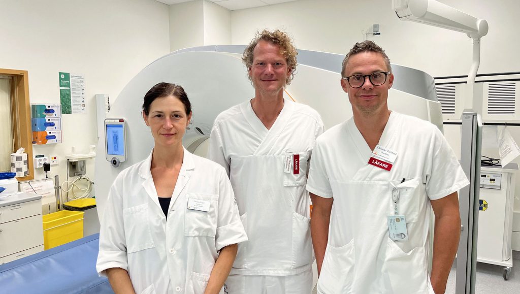
From the left: Viktoria Gaspar, Erik Thimansson, Christian Torbrand. Picture: Region Skåne.

Potential for a national work method—but one that requires everyone to move in the same direction

The working group has shared responsibility for driving the continued development and refinement of the diagnostic templates, but this doesn’t happen completely in isolation.

“We view ourselves as a hub that connects the requirements process,” Johan explains. “Our working group is made up of skilled specialists in their respective disciplines, and they have feelers out to the national working groups. For example, pathology has a quality and standardization committee for uropathology, where they discuss what should be in the template and link it to national care programs. Even outside the working group, work is done to ensure that the templates have the correct subject matter and incorporate medical relevance. Work is also being done to further adapt the solution to more technical standards such as openEHR and Snomed CT, and there is also a national working group involved in the process for those standards.”

“This is a bit difficult since everyone has their own opinions—radiologists are a bit like cats; everyone is strong-willed,” Erik says with a laugh. “I believe in having a small national group that agrees on changes and additions, which are then rolled out systematically. The risk is that if you have too many changes and local adaptations, the whole thing will turn into a jumble and fall apart. If the template gets too complicated and takes more time to use, it will no longer have any benefit in the end. The elegant thing about this template is that it’s simple. Of course, it’s always possible to supplement it with additional information in a free text field for people who feel they need that.”

After the successful pilot in Helsingborg, a rollout across all of Region Skåne is expected to take place in early 2024. This is a large region that will rapidly produce greater amounts of data. At the same time, the working group is conducting a national survey to identify other Swedish regions with similar conditions, i.e., where both radiologists and pathologists are already working in a digital diagnostic environment and registering their cases in INCA. They are already seeing high demand from users around the entire country.

“The first step will be to roll this out to all of Region Skåne, but we are aiming for this to become a national work method, so we’ll also work harder to bring about more widespread implementation in 2024,” Johan says.

“I see the potential for it to become a national solution, but that requires radiologists, pathologists, and urologists to all move in the same direction and requires the regions to do so as well, regardless of which IT solutions they use for diagnostics and reporting. We hope that in the future this feedback loop can be utilized by as many radiologists as possible nationwide,” Erik concludes.


References

Featured products & services