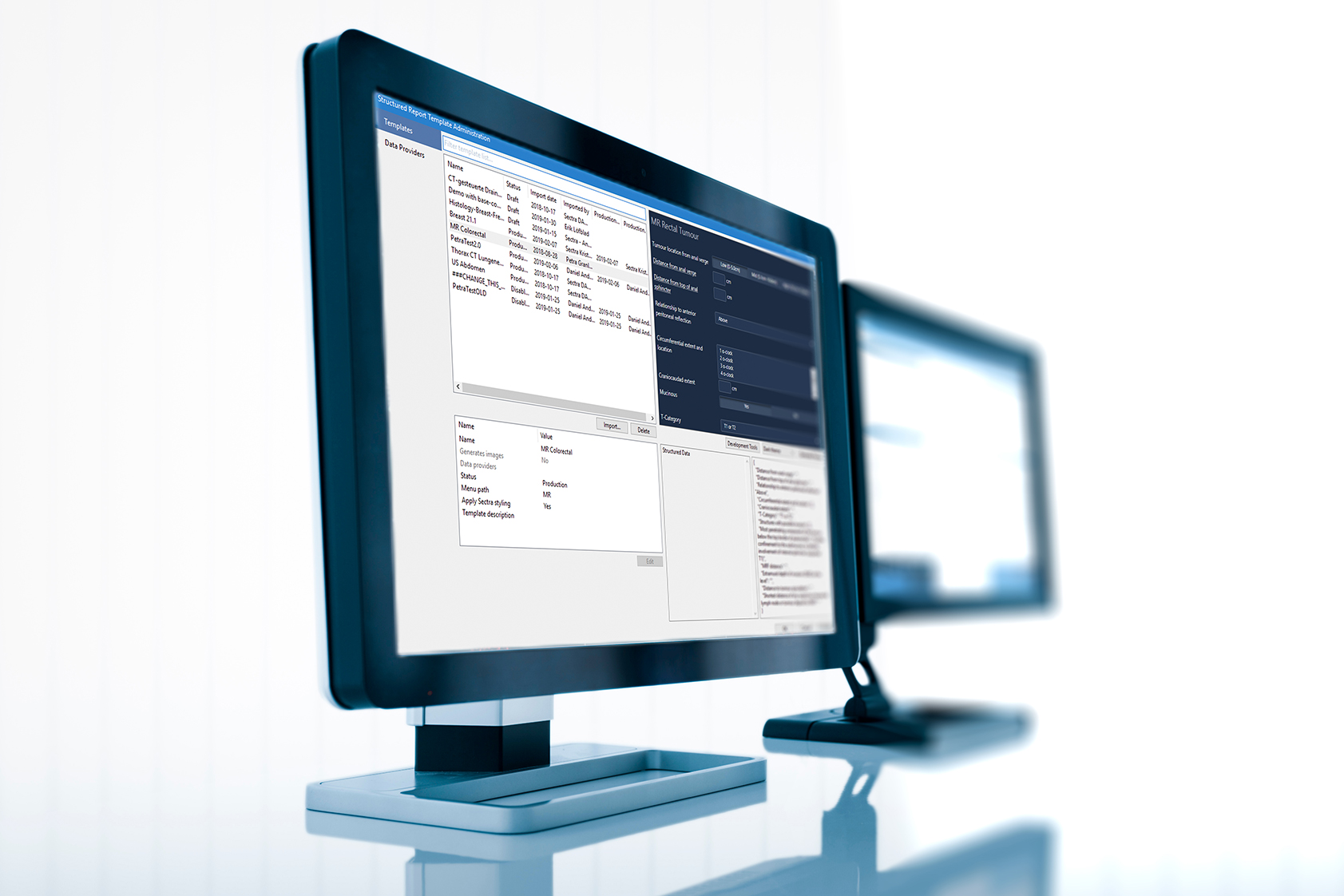
In 2020, Sectra and Sweden’s Regional Cancer Centre West (RCC) started working on a joint pilot project for MRI prostate reporting to improve and streamline data collection for individual patient overview (IPO) and national quality registers. The project was later successfully piloted with Region Gotland in spring 2021.
Leading the project with their expertise were Fredrik Jäderling, a radiology specialist at Capio St Göran’s Hospital in Stockholm, and Magnus Törnblom, a urology specialist at Visby Hospital. Fredrik, Magnus and Sectra worked together to develop a template for the Sectra PACS structured reporting framework as well as an integration with the RCC.
Many people have been involved from all different professions: urology, radiology, pathology, system developers, doctors, and nurses. It’s been a fantastic team effort—something I would like to see more of in healthcare. There are huge opportunities if we can work together in this way. I believe in digital templates instead of blocks of text that are impossible to evaluate.
“Prostate cancer is one of the most common cancers in Sweden and accounts for over 30 percent of cancer cases in men—every year, over 10,000 men are diagnosed,” Magnus says, adding that although prostate cancer accounts for the majority of cancer deaths among men, it can be cured if detected at an early, localized stage. “The big concern is that prostate cancer very often occurs in a benign form that grows very slowly and does not require treatment,” he continues. “When you screen for prostate cancer, you are very likely to encounter this form if you don’t really know where to look. This is known as overdiagnosis, which we want to avoid because it results in underdiagnosis of the aggressive form of the cancer that we actually want to find.”
Technology leap makes it possible to avoid over- and underdiagnosis
A major breakthrough in prostate cancer diagnosis came approximately six years ago, when biparametric magnetic resonance imaging (bpMRI) was introduced. With this special type of MRI, areas where aggressive prostate cancer is suspected can be identified. These can then be tested in more detail with targeted biopsies. The technique has revolutionized prostate cancer diagnosis and seems to largely address the problems of both over- and underdiagnosis.
“A major challenge, however, is that the MRI images are complex and difficult to interpret and radiologists today are generally unable to get feedback on what the tissue samples later showed,” Magnus explains. “As urologists, we are completely dependent on being able to trust what is written in the MRI report.”
“As a result, those of us working with IPO for prostate cancer, which is part of the national prostate cancer registry, developed a structured, digital reporting template for prostate MRIs with the help of the RCC and Fredrik Jäderling. Pathologists have also made a corresponding digital template for subsequent prostate biopsy results. The reporting templates are accepted nationally and, in addition to facilitating evaluations, also enable radiologists to get feedback from the biopsy results.”
The importance of structured reporting
A general concern is that medical records are difficult to understand and contain large amounts of unformatted text. “This makes it difficult to get an overview of the patient’s status, make compilations and evaluate what has been done at a group level,” Magnus says. “There is a need for additional system support where findings in medical images can be documented, followed up and reported in a structured way.”
“When we received a proposal to work with the RCC and the very committed Fredrik Jäderling, we were eager to help,” says Fredrik Lysholm at Sectra. “We already had a technical platform that we had made good progress on, but we lacked a clinical implementation of an overall workflow where we could demonstrate the path from report writing to distribution of structured information.”
Significant benefits from information exchange
The decision to focus the pilot project on prostate cancer diagnosis was based on the fact that it was already a well-established IPO diagnosis, where the overall benefit of information exchange between the urologist and pathologist is considerable. In autumn 2020, development of a reporting template for Sectra PACS began, supported by the model already developed within IPO.
There are a lot of gains you can make by registering MRI data as well as variables that we think are important for us to be able to follow up on the patient correctly. The link between the changes that the radiologist describes in the report and histological findings from the targeted biopsies that follow is an important feedback loop for improvement.
“In early March 2021, we were able to initiate a pilot in which Region Gotland started using the template in its reporting, thereby generating the structured information needed to send to the IPO and the National Prostate Cancer Registry (NPCR),” Fredrik Lysholm explains. “Once the staff had gotten used to it—and I felt that went very smoothly and quickly–the transfer of structured information to the RCC began. This also went well, and after monitoring the solution in practice for a number of weeks, we were able to compete the successful pilot just before the summer.”
Urologist Magnus Törnblom agrees that the project was very successful. He especially appreciates that the radiologist’s response in Sectra PACS is transferred to both the referring physician and the information network for cancer care (INCA). “I feel that there will be a clearer, more accurate, summarized assessment of the prostate in the structured response template than before,” he says.
Feedback flows between disciplines
Magnus further explains that now that Region Gotland has also started to register responses from pathologists via the INCA platform, he as a urologist receives complete information via IPO. This not only creates value for the patient and for him as a urologist, but also enables feedback flows between disciplines in the future so that healthcare staff can see the radiologist’s findings, where the biopsies were taken, and how the pathologist assessed the subsequent biopsies.
Radiologist Fredrik Jäderling, who was part of a development team when the reporting template for MR prostate was created, says, “There are a lot of gains you can make by registering MRI data as well as variables that we think are important for us to be able to follow up on the patient correctly. The link between the changes that the radiologist describes in the report and histological findings from the targeted biopsies that follow is an important feedback loop for improvement.”
A reporting template creates “huge opportunities”
In addition, everyone involved can get feedback on the biopsies ordered. Radiologist Fredrik Jäderling also believes that the advantage of this reporting template is that you must answer all the questions in it, which means that you do not omit any information, and the answers are always presented to the recipient in the same way. Like a guide, so you don’t forget anything.
“I think Sectra has been exemplary in engaging key people in the development of this MRI response template,” says Magnus Törnblom. “Many people have been involved from all different professions: urology, radiology, pathology, system developers, doctors and nurses. It’s been a fantastic team effort—something I would like to see more of in healthcare. There are huge opportunities if we can work together in this way. I believe in digital templates instead of blocks of text that are impossible to evaluate.”
Related cases


