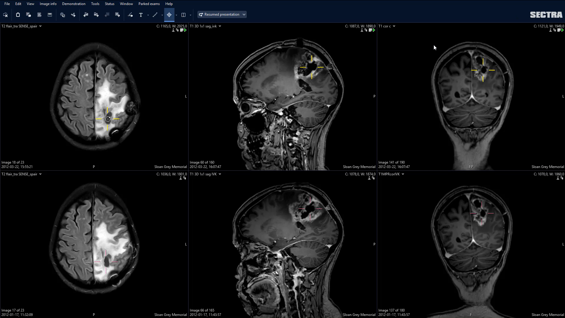
The Sectra Anatomical Linking tool
SAL is a fully automated tool for multi-modal rigid image registration—also referred to as anatomical linking—integrated with the Sectra PACS. It aims to increase reading efficiency by finding and synchronizing points of interest in CT, MR and nuclear medicine images, regardless of the frame of reference, slice thickness and field of view (FOV). It allows radiologists to automatically locate the same position in different images based on anatomical landmarks and thereby quickly identify and compare such findings as tumors and hemorrhages.
Study background
For a radiologist, the comparison of current and prior examinations is vital to accurately assess disease progression through the detection of sometimes very subtle changes that occur over time. However, the prior examinations create an additional workload for the radiologist interpreting a case, since the radiologist needs to review not only the current examination but also any prior examinations, which often requires a manual synchronization of images.
To assist radiologists in their interpretation of cases including one or more prior examinations, there are tools for synchronized image registration available that have been suggested to save time.
These “smart” applications are intended to enable a more efficient and effective comparison of the current examination with any prior examinations by performing a geometrical transformation between two image sets, i.e. an automatic spatial synchronization between images based on image similarities.
Method
Six radiologists (whereof three fellows) were recruited to read a set of cases (either 16 neuro or 14 musculoskeletal, MSK, cases) during two crossover reading sessions with at least four weeks in between sessions.
Each radiologist read each case twice, one time with synchronized navigation, which enables spatial synchronization across examinations from different study dates, and one time without. Both reading and reporting were performed using the Sectra PACS with synchronized navigation enabled by the integrated SAL functionality.
For each case, the time to complete the case was recorded, along with all PACS commands issued by the radiologist during the interpretation. The total efficiency was evaluated using the Wilcoxon signed-rank test.
After the second reading session, a questionnaire was distributed to each radiologist to assess their own impressions.
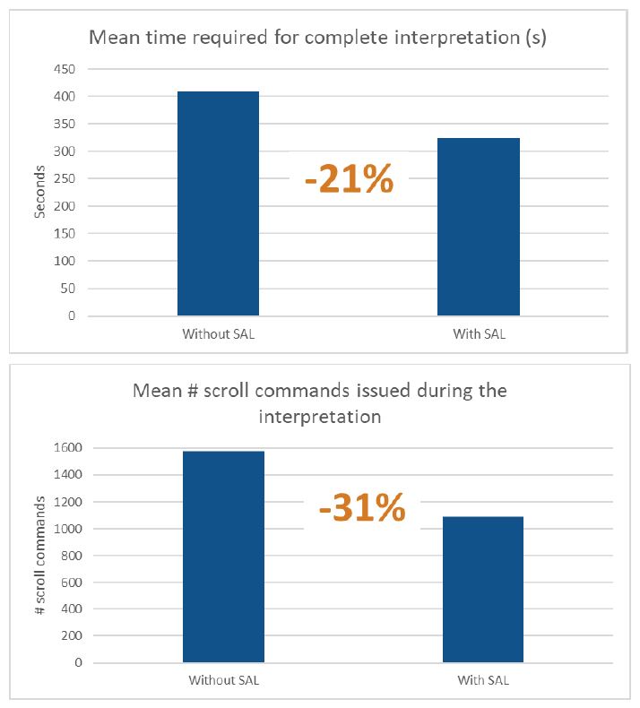
Result charts: Using the SAL functionality increased the efficiency of image interpretation by 21% and reduced the number of scroll commands by 31% in comparison to manual image synchronization.
Results
Significant improvements in efficiency were achieved using SAL, with an overall decreased reading time of 85 s (21%) and a decreased stack navigation of 484 scroll commands (31%).
The main conclusion is that automated image registration providing synchronized navigation across examinations from different study dates, such as SAL, enables radiologists to work more efficiently while reading cases with one or more prior examinations.
According to the results from the questionnaire (see figure below), the radiologists’ impressions of working with SAL reconfirm the efficiency benefits.
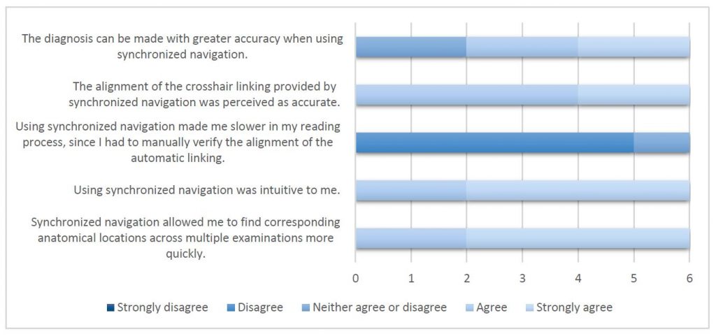
Results from the survey sent out to the radiologists after the second read to evaluate their impression of SAL.
Discussion
The statistical results confirm that synchronized navigation provides a significant improvement in interpretation efficiency both for time to interpret and the amount of scrolling while reading cases with one or more prior examinations.
It is worth noting that the mean improvement in time to interpret is somewhat larger for the neuro cases (−108 s) than for the MSK cases (−59 s); however, the mean time to read the neuro cases is also larger (482 s compared to 327 s without synchronized navigation). In terms of relative improvement, the average improvement is approximately equal, 22 and 18% for neuro and MSK, respectively, which is of the same magnitude as the overall average relative improvement of 21%.
References
Forsberg, D., Gupta, A., Mills, C., MacAdam, B., Rosipko, B., Bangert, B. A., . . . Sunshine, J. L. (2016). Synchronized navigation of current and prior studies using image. 12(3).
Note: The paper was written in collaboration between CMIV (Center for Medical Image Science and Visualization) and University Hospitals of Cleveland and published in the International Journal of Computer Assisted Radiology and Surgery in 2016.