Case description
During the aftermath of an ischemic stroke, a local swelling of the brain tissue develops—and there is a risk of brain herniation due to the expansive effect. When it involves the territory of the middle cerebral artery (MCA), it is termed malignant MCA infarction. Decompressive hemicraniectomy can be a lifesaving procedure, and it is most effective when performed early. The prediction of a malignant development of the MCA infarction is best accomplished by measuring the volume of damaged brain tissue.
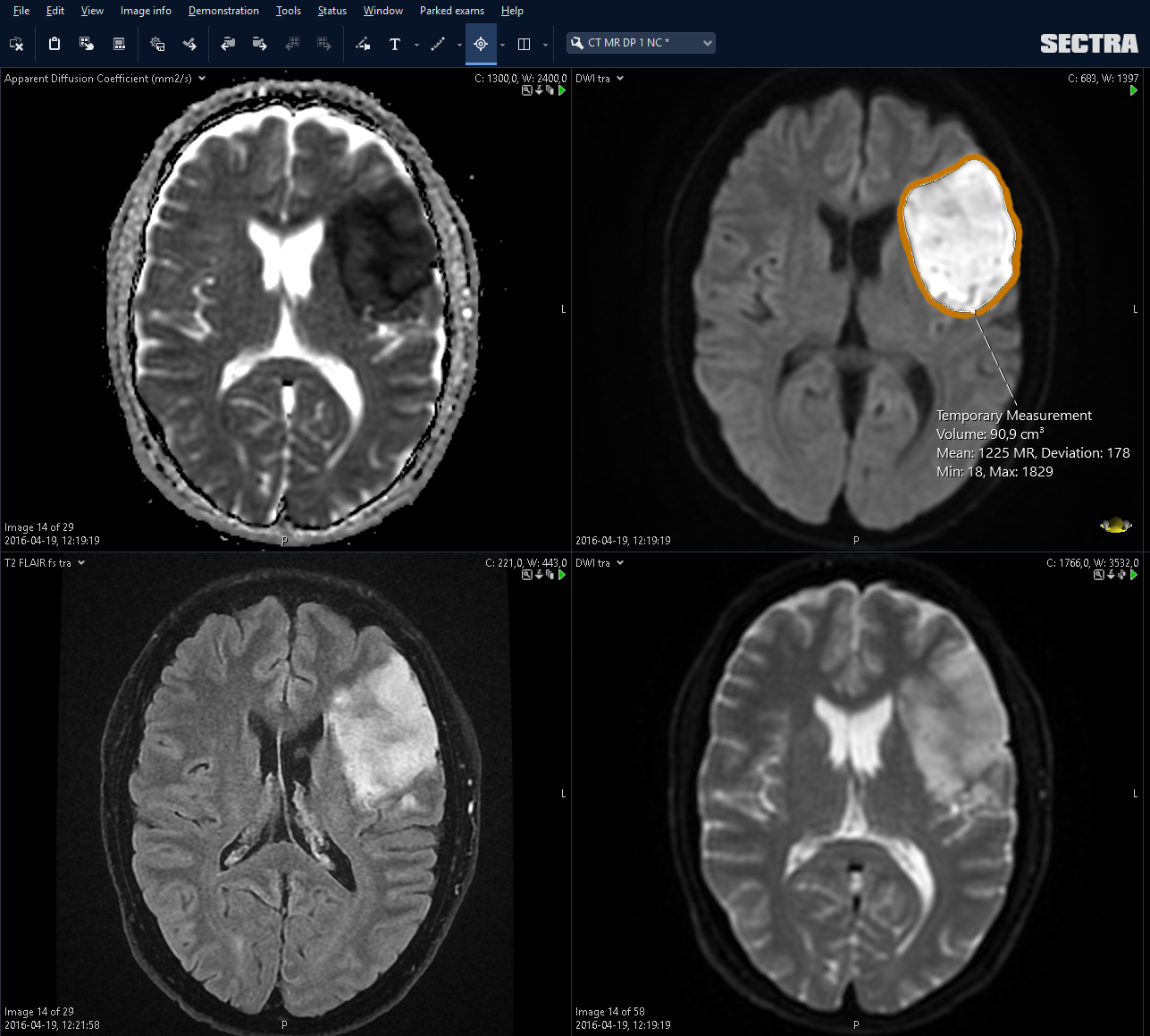
The study performed at the Department of Neuroradiology at Skåne University Hospital in Lund, Sweden, aimed to examine how well three different volume measurement tools measure the volume of acute cerebral infarcts on diffusion-weighted MRIs.
Method
Two independent reviewers performed volume measurements of acute infarctions on diffusion-weighted MRIs of 27 patients. They used three different methods in the study:
- Manual volume measurement (reference method)
- Semi-automatic measurement
- Automatic measurement (the Sectra Volume Measurement tool)
The obtained volume and time of measurement were registered.
Results
The differences in volume using the three different measurement methods were very small and not statistically significant. The different measurement methods were thus concluded not to have any clinically relevant impact with respect to volume.
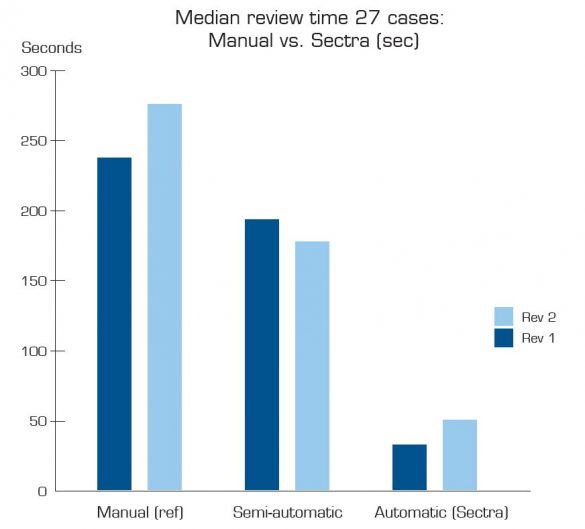
The median review time for the two reviewers was 237 and 275 seconds, respectively, using the manual method, 193 and 177 seconds using the semi-automatic method, and 32 and 50 seconds using the automatic method.

Discussion
This study shows that the three methods are equally able to determine the infarct volume and that the automatic method is much faster than the manual and semiautomatic methods.
Automatic volume segmentation is the best and fastest method of performing volume measurements of acute infarctions on diffusion-weighted MRIs in clinical routine practice.
Study authors
Kristina Galic and Sofia Patarroyo.
Supervisor: Johan Wassélius, MD, PhD.
Bibliography
1. Galic, Kristina och Patarroyo, Sofia. Volymmätning av cerebrala infarkter med tre olika MR-baserade metoder – vilken är bäst? Lund, Sweden : Skåne University Hospital, 2017.
* Volume measurement of damaged brain tissue of the middle cerebral artery on diffusion-weighted magnetic resonance images (MRIs) for ischemic stroke patients.


