Let’s talk
Interested to learn more, or to schedule a demo? Don’t hesitate to get in touch.
4 × Study
With the right smart and fast clinical tools, relevant time savings can be achieved
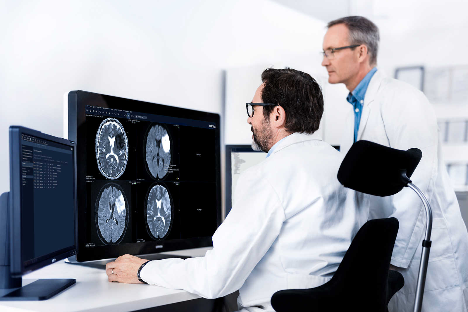
With steadily growing patient exams and increasingly complex and time-critical cases, radiology reading efficiency and the ability to communicate actionable reports are key for patient outcomes. With the right smart and fast clinical tools, relevant time savings can be achieved. See how four different clinical tools from Sectra — Volume Measurement, Lesion Tracking, Vessel Analysis and Anatomical Linking — have proven to provide significant time savings, boost efficiency and increase the quality in radiologists’ reviews.
Decompressive hemicraniectomy can be a lifesaving procedure and is most effective when performed early. The prediction of a malignant development of the middle cerebral artery (MCA) infarction is best accomplished by measuring the volume of damaged brain tissue.
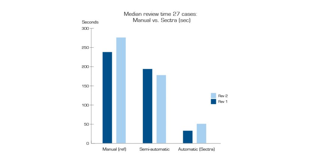
This study aimed to examine how well three different methods measured the volume of acute cerebral infarcts on diffusion-weighted MRIs. The automatic Sectra Volume Measurement tool was determined to be statistically significantly faster than manual volume measurements. Of the 27 patient cases studied, the two reviewers saved 3 minutes 25 seconds and 3 minutes 45 seconds, respectively, on the median case compared with the manual estimation. Sectra’s tool was proven to be 7.4 times and 5.5 times faster, respectively, than the manual method.
At the Oslo University Hospital, radiology capacity has been identified as a bottleneck, especially when it comes to CT and MRI case reviews with the high number of images and previous exams requiring timeconsuming comparisons. This study aimed to investigate whether Sectra Lesion Tracking (SLT) could raise efficiency in measuring, tagging and comparing lesions on CT and MRI exams for complex cancer cases. It also focused on creating a business case to guide a potential future purchasing decision concerning SLT.
For reviewed CT exams (from initial review to dictation) of complex cancer cases, the average time saved using the SLT tool was 15.4 minutes per case (corresponding to a 35% reduction in time), and an additional 3 minutes was saved when measured from review to sign-off. The authors concluded that the time savings were higher than expected. SLT resulted, “without a doubt”, in increased efficiency in reviews as well as better diagnostic quality.
The comparison of current and prior examinations is vital to accurately assess disease progression through the detection of sometimes very subtle changes that occur over time. However, the prior examinations create an additional workload for the radiologist interpreting a case, which often requires a manual synchronization of images. To assist radiologists in their interpretation of cases including one or more prior examinations, there are tools for synchronized image registration available that have been suggested to save time.
This study aimed to evaluate the time savings of using the Sectra Anatomical Linking (SAL) application, an automated tool for multi-modal rigid image registration integrated within the Sectra PACS. The results showed significant improvements, with an overall decreased reading time of 85 s (21%) and a decreased stack navigation of 484 scroll commands (31%).
The calculation of the degree of carotid artery stenosis is an important component in the diagnosis of stroke, especially for patients with significant stenosis (>50%). This study aimed to investigate the time savings using a semi-automated stenosis measurement tool on stroke patients compared to manual measurement.
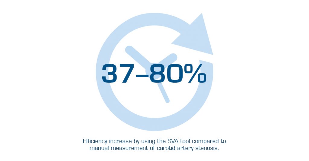
The study resulted in the conclusion that radiologists perform faster calculations using Sectra Vessel Analysis (SVA) compared to manual measurement of carotid artery stenosis. The time savings varied from 20 seconds (37% efficiency increase) to 132 seconds (80% efficiency increase). The average time savings was 68 seconds, equivalent to a 53% increase in efficiency.
The main benefits of SVA included ease of use, speed, and increased accuracy in the measurement of diameter when the carotid artery was curved. It also increased radiologist confidence in measurement accuracy and overall assessment.
Interested to learn more, or to schedule a demo? Don’t hesitate to get in touch.


